. 2014 May 8;157(4):910-21.
doi: 10.1016/j.cell.2014.03.037.

Enhanced protein degradation by branched ubiquitin chains

Affiliations

Enhanced protein degradation by branched ubiquitin chains

Hermann-Josef Meyer et al. Cell. .

Abstract

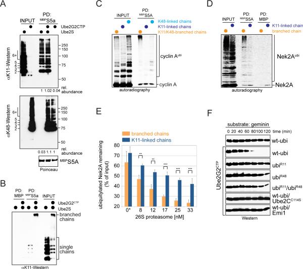
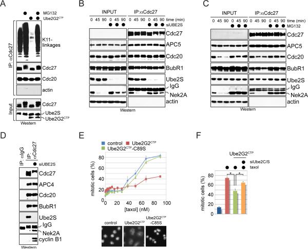
Posttranslational modification of cell-cycle regulators with ubiquitin chains is essential for eukaryotic cell division. Such chains can be connected through seven lysine residues or the amino terminus of ubiquitin, thereby allowing the assembly of eight homogenous and multiple mixed or branched conjugates. Although functions of homogenous chain types have been described, physiological roles of branched structures are unknown. Here, we report that the anaphase-promoting complex (APC/C) efficiently synthesizes branched conjugates that contain multiple blocks of K11-linked chains. Compared to homogenous chains, the branched conjugates assembled by the APC/C strongly enhance substrate recognition by the proteasome, thereby driving degradation of cell-cycle regulators during early mitosis. Our work, therefore, identifies an enzyme and substrates for modification with branched ubiquitin chains and points to an important role of these conjugates in providing an improved signal for proteasomal degradation.


Figures

Figure 1
Figure 1. Ube2S is required for APC/C-substrate ubiquitylation in prometaphase
A. Ube2S was depleted from prometaphase HeLa cells resistant to hygromycin (HeLa-HR) or stably expressing siRNA-resistant Ube2S, and analyzed for Nek2A-levels by Western. B. HeLa cells transfected with control or Ube2S-siRNAs were synchronized in prometaphase, treated with cycloheximide, and analyzed for protein abundance by Western. C. HeLa cells expressing Hisubiquitin and transfected with control- or Ube2S-siRNA were synchronized in prometaphase and treated with MG132. Purified ubiquitin conjugates were analyzed by αNek2A- or αCdc20-Western. D. Nocodazole-arrested HeLa cells treated with control- or Ube2S-siRNAs were released into drug-free medium and protein levels were analyzed by Western. E. Depletion of Ube2S delays anaphase initiation. HeLa cells stably expressing GFPtubulin and mCherryhistone H2B were transfected with control- or Ube2S-siRNA and monitored by live-cell imaging. The time from nuclear envelope breakdown to initiation of sister chromatid separation was determined for >130 cells per condition (n=3). See also Figure S1.
Figure 2
Figure 2. The APC/C assembles branched ubiquitin chains
A. Nek2A is modified with branched chains in vitro. 35S-labeled Nek2A272-445 was incubated with APC/CCdc20, Ube2C, Ube2S, and ubiquitin mutants. Reactions were analyzed by autoradiography. B. 35S-labeled Ub-cyclin A was incubated with APC/CCdc20, Ube2S, and ubiquitin mutants. Reactions were analyzed by autoradiography. C. Ub-cyclin A, UbR11-cyclinA, Ub-Ubcyclin A and UbR11-Ub-cyclin A were incubated with APC/C, Ube2S, and methylubiquitin or ubiquitin. Left: reaction products if Ube2S would elongate or branch chains (pink: fused substrate-ubiquitin; red: ubiquitin added by Ube2S). Right: Reactions were analyzed by αHA-Western. D. Ub-cyclin AHA, Ub-Ub-cyclin AHA, or Ub-Ub-Ub-cyclin AHA were incubated with APC/CCdh1, Ube2S and methylubiquitin or ubiR11. Reaction products were analyzed by αHA-Western. Red dot: first added ubiquitin; blue dot: second added ubiquitin; green dot: third added ubiquitin. E. Ube2S efficiently modifies Nek2A with branched chains in vitro. Ub3-Nek2A was incubated with APC/C, Ube2S, and methylubiquitin or ubiR11, and analyzed by αNek2A-Western. See also Figure S2.
Figure 3
Figure 3. The APC/C assembles branched chains in cells
A. Outline of a method to monitor synthesis of branched chains. A TEV-site and FLAG-epitope (ubiTEV/FLAG) were introduced into ubiquitin. Cleavage of ubiTEV/FLAG-conjugates reveals a ~2kDa stamp for elongation or multiple stamps for branching. B. ubiTEV/FLAG is recognized by Ube2S and APC/C and sensitive to TEV cleavage. Ub-cyclin AHA was incubated with APC/C, Ube2S, and wt-ubiquitin or ubiTEV mutants. Reactions were treated with TEV and analyzed by αHA-Western. C. 35S-cyclin A was incubated with APC/C, Ube2C, Ube2S, and ubiquitin, ubiTEV or ubiTEV/FLAG proteins. Reactions were treated with TEV and analyzed by autoradiography. D. Ub-cyclin A or Ub-Nek2A272-445 were incubated with APC/C, Ube2S, ubi53TEV and ubi64TEV/FLAG to add unbranched K11-linked chains; alternatively, cyclin A or Nek2A were modified by APC/C, Ube2C, Ube2S, ubi53TEV and ubi64TEV/FLAG with branched chains. Reactions were treated with TEV and analyzed by αFLAG-Western. E. Nek2A272-445 was incubated with APC/C, Ube2C, Ube2S, ubi53TEV and ubi64TEV/FLAG. APC/C was purified from G1 phase, when it is active, or S phase, when APC/C-activity is low. APC/C was also inhibited by Emi1. Reactions were treated with TEV and analyzed by αFLAG-Western. F. APC/C was purified from prometaphase HeLa cells transfected with control-or Ube2S-siRNA and analyzed for co-purifying K11-linked chains using αK11-Western. G. APC/C synthesizes branched chains in vivo. APC/C was purified from prometaphase cells expressing FLAGubi53TEV and Hisubi64TEV/FLAG, treated with TEV, and analyzed by αFLAG-Western. Upper panel: conjugates co-purifying with APC/C. Lower panel: MW-range for diagnostic peptides (longer exposure). Ubiquitin detected in reactions without TEV is produced by DUBs that co-precipitate with APC/C. H. APC/C was purified from cells WT- or Lys-mutant FLAGubi53TEV and HISubi64TEV/FLAG, treated with TEV, and analyzed for co-purifying conjugates (upper panel) or diagnostic peptides (lower panel). I. Nek2A is modified with branched chains in cells. HANek2A was purified from cells expressing FLAGubi53TEV and Hisubi64TEV/FLAG and treated with TEV. Reactions were analyzed by αFLAG-Western. See also Figure S3.
Figure 4
Figure 4. Synthesis of branched chains with defined composition
A. 35S-cyclin A was incubated with APC/CCdh1 and no E2, Ube2G2, or Ube2G2CTP. Reactions were analyzed by autoradiography. B. 35S-labeled cyclin A was incubated with APC/CCdh1, low levels of Ube2C, and Ube2G2CTP or Ube2S. APC/C was inhibited by Emi1 or an excess of recombinant APC/C-substrate. Reactions were analyzed by autoradiography. C. 35S-geminin or mutants in its D-box or initiation motif (IM) were incubated with APC/C, low levels of Ube2C, and Ube2G2CTP or Ube2S. Reactions were analyzed by autoradiography. D. Ube2G2CTP catalyzes chain formation with ubiK48. 35S-cyclin A was incubated with APC/C, low levels of Ube2C, Ube2S or Ube2G2CTP, and ubiquitin mutants. Reactions were analyzed by autoradiography. E. Ube2G2CTP requires K48 of ubiquitin for chain formation. 35S-cyclin A was incubated with APC/C, low levels of Ube2C, Ube2S or Ube2G2CTP, and ubiquitin variants. Reactions were analyzed by autoradiography. F. Ube2G2CTP catalyzes K48-linked chain formation with APC/C. Ub-cyclin A was incubated with APC/C, either Ube2S or Ube2G2CTP, and ubiquitin. Reaction products were analyzed by Western. G. Ube2S and Ube2G2CTP synthesize high-MW conjugates. Ubcyclin A was mixed with Ube2G2CTP and low levels of ubiquitin, which leads to addition of two K48-linked ubiquitin molecules (Ub3-cyclin A). Next, an excess of ubiR48 and Ube2S were added. Ub-cyclin A was also directly incubated with Ube2S and ubiR48. Reactions were analyzed by Western. H. High-MW chains are seen in single substrate-turnover assays. An excess of Ub-cyclin A (1μM) over APC/CCdh1 (~10nM) was incubated with Ube2S, Ube2S/Ube2G2CTP, or Ube2S and Ube2G2CTP-C89S. Reactions were analyzed by Western. I. High-MW conjugates assembled by Ube2S and Ube2G2CTP are branched chains. Ub-cyclin A was incubated with APC/C, Ube2S, Ube2G2CTP, and ubiquitin mutants under single substrate-turnover conditions and analyzed by Western. Mixtures of ubiR11 and ubiR48 allow Ube2S and Ube2G2CTP to synthesize mixed chains, yet prevent introduction of branches. See also Figures S4 and S5.
Figure 5
Figure 5. Branched chains enhance proteasomal degradation
A. Ub-cyclin A was modified by the APC/C and Ube2S or Ube2G2CTP with either K11- or K48-linked chains. Reaction products were incubated with immobilized MBPS5a either alone or together, and analyzed by Western. B. Proteins decorated with K11/K48-linked branched chains bind more efficiently to S5a than those modified with single chains. Ub-cyclin A modified with K11-linked or K11/K48-branched chains were incubated with MBPS5a under stringent conditions, and binding was monitored by Western. C. Preferential binding of branched chains is independent of chain length. 35S-labeled cyclin A was modified by APC/C, low levels of Ube2C, Ube2S and/or Ube2G2CTP, and ubiquitin, ubiK11, or ubiK48 to assemble K11-, K48-linked, or K11/K48-branched chains, and incubated with S5a. D. Branched chains increase the affinity of Nek2A for the proteasomal receptor S5a. 35S-labeled Nek2A272-445 was modified by APC/C, Ube2C and Ube2S with homogenous or branched chains (using ubiK11 or wild-type ubiquitin) and incubated with MBPS5a. Reactions were analyzed by autoradiography. E. Branched chains improve the efficiency of proteasomal degradation in vitro. 35S-HANek2A272-445 was modified with branched or homogenous K11-linked chains and added to increasing concentrations of 26S proteasomes. Five independent titrations were monitored by autoradiography and quantified using ImageJ (n=5; data are average +/- SD; ** p<0.002 in paired t-test; *** p<0.001). The asterisk marks background degradation by proteasomes that co-purify with APC/C. F. Branched chains promote degradation more efficiently. Prometaphase extracts with endogenous Ube2S were treated with Ube2G2CTP in the presence of ubiquitin mutants. The stability of geminin was monitored by Western. Geminin was stable in extracts containing mixtures of ubiR11 and ubiR48, a condition that allows Ube2S and Ube2G2CTP to assemble mixed, but not branched chains. See also Figure S6.
Figure 6
Figure 6. Branched chains promote proteasomal degradation in cells
A. Branched chains facilitate extraction of ubiquitylated substrates from the APC/C. HeLa cells stably expressing Ube2G2CTP and endogenous Ube2S were released from nocodazole-induced mitotic arrest and treated with proteasome inhibitor MG132, as indicated. Co-purifying K11-conjugates were monitored by Western. B. Branched chains facilitate extraction of Nek2A from the APC/C. HeLa cells transfected with control- or Ube2S-siRNA were synchronized in prometaphase and treated with cycloheximide. APC/C was purified at indicated times after cycloheximide addition, and co-purifying proteins were analyzed by Western. C. The stability of Nek2A-binding to the APC/C was analyzed in prometaphase HeLa cells treated with cycloheximide and proteasome inhibitors. D. APC/C was purified from prometaphase HeLa cells treated with control- or Ube2S-siRNA, and levels of co-purifying proteins were determined by Western. E. Stable expression of Ube2G2CTP, but not Ube2G2CTP-C89S, causes spindle checkpoint bypass. Upper panel: HeLa cells stably expressing indicated proteins were treated with taxol. Mitotic arrest was scored by automated microscopy (n=3; >1000 cells/condition; data are average +/- SD; unpaired t-tests p<0.0005). Lower panel: Images of representative cells stained with Hoechst. F. Checkpoint bypass requires Ube2G2CTP and Ube2C/Ube2S. HeLa cells or stable cell lines expressing Ube2G2CTP were treated with siRNAs against Ube2C and Ube2S as well as taxol, and the mitotic index was determined by automated microscopy (n=3; 1000 cells/condition; data are average +SD; paired t-test p<0.02). See also Figure S7.
Figure 7
Figure 7. Enhanced protein degradation by branched ubiquitin chains
APC/C-substrates are initially modified by Ube2C with short chains containing K11-, K48-, and K63-linkages. Ube2S adds blocks of ~6 K11-linked ubiquitin molecules to each chain moiety with an unmodified K11. This process leads to the formation of branched ubiquitin chains that increase the proteolytic capacity of the initial ubiquitylation mark.

Comment in

Similar articles

Cited by

References

    1. Amador V, Ge S, Santamaria PG, Guardavaccaro D, Pagano M. APC/C(Cdc20) controls the ubiquitin-mediated degradation of p21 in prometaphase. Molecular cell. 2007;27:462–473. - PMC - PubMed
    1. Boname JM, Thomas M, Stagg HR, Xu P, Peng J, Lehner PJ. Efficient internalization of MHC I requires lysine-11 and lysine-63 mixed linkage polyubiquitin chains. Traffic. 2010;11:210–220. - PMC - PubMed
    1. Chau V, Tobias JW, Bachmair A, Marriott D, Ecker DJ, Gonda DK, Varshavsky A. A multiubiquitin chain is confined to specific lysine in a targeted short-lived protein. Science. 1989;243:1576–1583. - PubMed
    1. Dammer EB, Na CH, Xu P, Seyfried NT, Duong DM, Cheng D, Gearing M, Rees H, Lah JJ, Levey AI, et al. Polyubiquitin linkage profiles in three models of proteolytic stress suggest the etiology of Alzheimer disease. J Biol Chem. 2011;286:10457–10465. - PMC - PubMed
    1. Deshaies RJ, Joazeiro CA. RING domain E3 ubiquitin ligases. Annu Rev Biochem. 2009;78:399–434. - PubMed

Publication types

MeSH terms

Substances