. 2014 May 8;157(4):935-49.
doi: 10.1016/j.cell.2014.02.057.

Transit-amplifying cells orchestrate stem cell activity and tissue regeneration

Affiliations

Transit-amplifying cells orchestrate stem cell activity and tissue regeneration

Ya-Chieh Hsu et al. Cell. .

Abstract

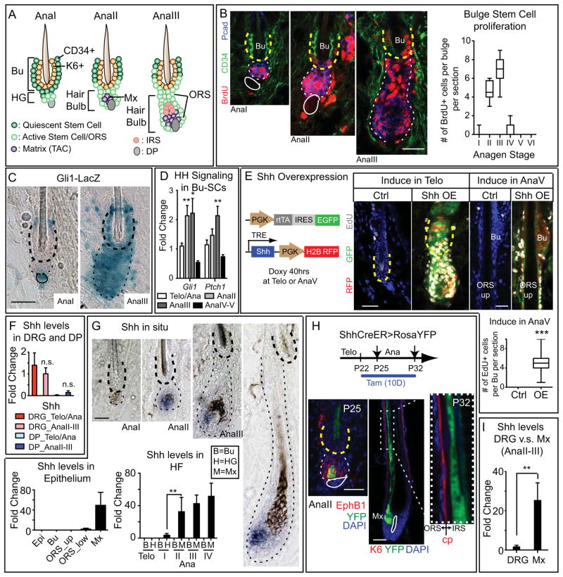
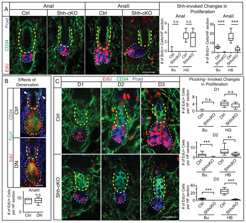
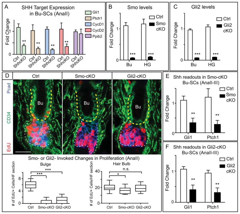
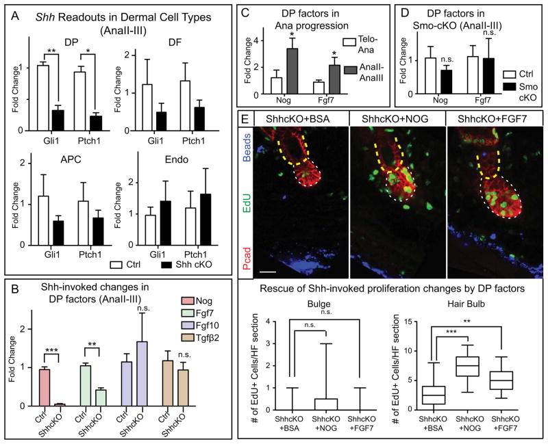
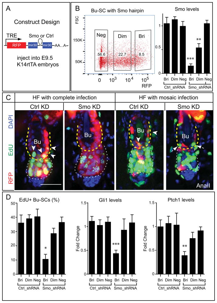
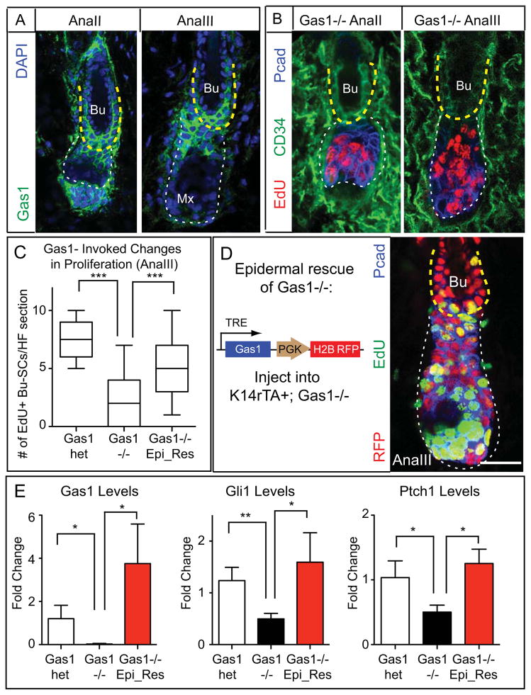
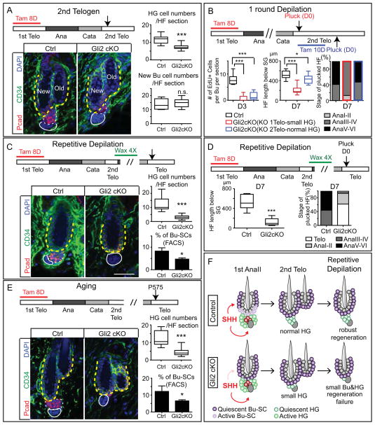
Transit-amplifying cells (TACs) are an early intermediate in tissue regeneration. Here, using hair follicles (HFs) as a paradigm, we show that emerging TACs constitute a signaling center that orchestrates tissue growth. Whereas primed stem cells (SCs) generate TACs, quiescent SCs only proliferate after TACs form and begin expressing Sonic Hedgehog (SHH). TAC generation is independent of autocrine SHH, but the TAC pool wanes if they can't produce SHH. We trace this paradox to two direct actions of SHH: promoting quiescent-SC proliferation and regulating dermal factors that stoke TAC expansion. Ingrained within quiescent SCs' special sensitivity to SHH signaling is their high expression of GAS1. Without sufficient input from quiescent SCs, replenishment of primed SCs for the next hair cycle is compromised, delaying regeneration and eventually leading to regeneration failure. Our findings unveil TACs as transient but indispensable integrators of SC niche components and reveal an intriguing interdependency of primed and quiescent SC populations on tissue regeneration.


Figures

Figure 1
Figure 1. HH Pathway Activity in Bu-SC is Up-regulated upon Bu-SC Activation Concurrent with Matrix SHH Expression, while SHH Overexpression Induces Bu-SC Proliferation
(A) Schematics of early anagen from AnaI-III. The AnaII hair bulb contains newly emerged matrix (Mx). From AnaIII and onward, the HG structure is no longer obvious. (B) Time-course monitoring of BrdU+ cells during anagen and quantifications. Bulge: CD34+ (in green) and yellow dashed lines. HG/hair bulb: Pcad+ (in blue) and white dashed lines. Solid lines, DP (≥ 30 HFs from 2–3 animals per substage). (C) β-galactosidase activity (blue) of Gli1-LacZ HFs at AnaI and AnaIII. Bulge, thick dashed lines; hair bulb, thin dashed lines; solid lines, DP. (D) RT-PCR of Gli1 and Ptch1 from purified Bu-SCs at different substages. (E) Schematics and results of Shh overexpression in telogen and AnaV compared to controls. (F) RT-PCR examining Shh expression in dissected DRGs and FACS-purified DP. (G) In situ hybridization (purple) and RT-PCR examining Shh levels in different compartments within epithelium. (H) Tamoxifen treated Shh-CreER>RosaYFP HFs at different anagen substages. EphrinB1 (EphB1) marks the hair bulb. Companion layer (Cp) is K6+ and is sandwiched between IRS and ORS. YFP+ cells are only seen in IRS not ORS. (I) RT-PCR of Shh from DRG and FACS-purified matrix. Data are mean±SD. *:p<0.05; **:p<0.01. ***:p<0.001. n.s.: not significant. Box-and-whisker plots: mid-line, median; Box, 25th and 75th percentiles; whiskers, minimum and maximum. Scale bars: 30μm.
Figure 2
Figure 2. TAC-Derived SHH is Essential for Bu-SC and Hair Bulb Proliferation During Normal and Plucking-Induced Hair Regeneration
(A) Conditional deletion of Shh from HFs impairs proliferation of both Bu-SCs and hair bulbs (n≥2 mice, ≥12 HFs per mice). (B) Immunolocalization of EdU, CD34, and TUJ1 (pan-neuronal marker which also marks IRS) in AnaIII HFs and quantifications of EdU+ cells per bulge in sections from control and denervated side (DN) of same animal (n=3 mice; ≥14 HFs per mice). Arrowheads: nerve fibers innervating the HF. (C) HFs from WT and Shh-cKO were plucked during telogen at D0, and the proliferative status of Bu-SCs and hair bulbs were examined at D1, D2 and D3 post plucking (n≥2 mice, ≥8 HFs per mice). Box-and-whisker plots as in Figure 1 legend. Scale bar: 30μm. n.s., not significant. **:p<0.01; ***:p<0.001. Bulge (Bu), yellow dashed lines; HG or hair bulb (HB), white dashed lines. Scale bars: 30μm. n.s., not significant.
Figure 3
Figure 3. SHH Pathway Activity in the HF is Critical for Bu-SC Activation
(A) RT-PCR of SHH targets in FACS-purified control or Shh-cKO Bu-SCs at AnaII. Known SHH targets are down-regulated and control gene Ppib2 is un-changed in Shh-cKO Bu-SCs. (B) RT-PCR of Smo in FACS-purified Bu-SCs and HG in control and Smo-cKO. (C) RT-PCR of Gli2 in FACS-purified Bu-SCs and HG in control and Gli2-cKO. (D) AnaII HFs from control, Smo-cKO, and Gli2-cKO, with immunolabeling of EdU, CD34, and Pcad. (n≥3 per genotype, ≥14 HFs per mouse). Bulge, yellow dashed lines; hair bulb, white dashed lines. Box-and-whisker plots as in Figure 1 legend. Scale bar: 30μm. (E–F) RT-PCR of Gli1 and Ptch1 in FACS-purified control, Smo-cKO, or Gli2-cKO Bu-SCs at AnaII-III. Data are mean±SD. n.s., not significant. *:p<0.05; **:p<0.01; ***:p<0.001.
Figure 4
Figure 4. TAC-Derived SHH Maintains the Expression of DP Activation Signals
(A) RT-PCR of Gli1 and Ptch1 in FACS-purified DP, DF, adipocyte precursor cells (APC) and endothelial cells (Endo) from control and Shh-cKO at AnaII-III. (B) RT-PCR of DP factors in FACS-purified control and Shh-cKO DP at AnaII-III. (C) RT-PCR of Nog and Fgf7 at telogen→anagen transition and at AnaII-III. (D) RT-PCR of Nog and Fgf7 in control and Smo-cKO DP at AnaII-III. (E) BSA, NOGGIN, or FGF7 was coinjected with Cy5 fluorescent beads (blue) into Shh-cKO skin for 3 days, followed by EdU administration at D3 and analysis at D4. (n=3 mice per experiment; ≥12 HFs per mouse). Bulge, yellow dashed lines; hair bulb, white dashed lines. Box-and-whisker plots as in Figure 1 legend. Scale bar: 30μm. Data are mean±SD. n.s., not significant. *:p<0.05; **:p<0.01; ***:p<0.001.
Figure 5
Figure 5. SHH is Required Autonomously in Bu-SCs for Their Activation
(A) Lentiviral construct design. (B) FACS plot of a mosaically transduced animal showing percentage of RFPbri (Bri), RFPdim (Dim), and RFPneg (Neg) Bu-SCs, and RT-PCR examining Smo from FACS-purified Bu-SCs infected with hairpins against luciferase (Ctrl_shRNA) or Smo (Smo_shRNA). (C) AnaII HFs either completely infected or mosaically infected by Ctrl or Smo hairpins, immunolabeled with RFP and EdU. Arrowheads mark RFP and EdU double positive Bu-SCs in HF infected with Ctrl_shRNA. Arrows mark EdU single-positive Bu-SCs in HF infected with Smo_shRNA. (D) Quantifications of EdU incorporation and RT-PCR of Gli1 and Ptch1 in Ctrl- or Smo- shRNA infected Bu-SCs (n=2 per hairpins). Bulge, yellow dashed lines; hair bulb, white dashed lines. Data are mean±SD. Scale bar: 30μm. *:p<0.05; **:p<0.01; ***:p<0.001.
Figure 6
Figure 6. GAS1 is Important for Proper Bu-SC Activation
(A) Immunodetection of GAS1. (B) Gas1−/− HFs immunolabeled with EdU, CD34 and Pcad. (C) Quantifications of EdU incorporation in AnaIII Bu-SCs from Gas1het, Gas1−/−, and Gas1−/− with epidermal rescue of GAS1 expression (n≥2 mice, ≥14 HFs per mouse). (D) (Left) Experimental strategy and lentiviral construct design. GAS1 was turned on by feeding the infected mice with Doxy chow 3 days prior to anagen entry until the time of analysis. (Right) AnaIII Gas1−/− HF with GAS1 expression restored in the epidermal compartment. Tissue is immunolabeled for RFP, EdU and P-cadherin. (E) RT-PCR examining levels of Gas1, Gli1, and Ptch1 in FACS-purified AnaIII Bu-SCs from Gas1het, Gas1−/−, and Gas1−/− with epidermal rescue of GAS1 expression. Bulge, yellow dashed lines; hair bulb, white dashed lines. Box-and-whisker plots as in Figure 1 legend. Scale bars: 30μm. Data are mean±SD. n.s., not significant. *:p<0.05; **:p<0.01; ***:p<0.001.
Figure 7
Figure 7. Defective Bu-SC Proliferation Leads to Reduced HG Cell Number Short-Term and Regeneration Failure Long-term
(A) HG cell numbers are reduced after Gli2-cKO HFs complete one cycle. Examination was in 2nd telogen (n≥3 mice, ≥18 HFs per mouse). (B) Control and Gli2-cKO mice following one round of hair plucking. Gli2 is knocked-out either in 1st telogen (red bars) or 2nd telogen (blue bars). Plucking was performed in 2nd telogen. HFs were examined at D3 for Bu-SCs proliferation and at D7 for HF length [below sebaceous gland (SG)] and hair cycle stage (n≥2 mice, ≥8 HFs per mouse). (C) Control and Gli2-cKO HF after 4 waxings (n≥3 mice, ≥21 HFs per mouse). (D) Control and Gli2-cKO HFs were plucked after 4 waxings and examined at D7 for HF length (below SG) and hair cycle stages (n≥2 mice, ≥9 HF per mice). (E) Aged control and Gli2-cKO HFs and quantifications. (F) Model summarizing the results. Bulge: CD34+ (in green) and yellow dashed lines. HG: Pcad+ (in red) and white dashed lines. Data are mean±SD. *:p<0.05; ***:p<0.001. n.s., not significant. Box-and-whisker plots as in Figure 1 legend. Scale bars: 30μm.

Comment in

  • Tic-TACs: refreshing hair growth.
    Kalaitzidis D, Scadden DT. Kalaitzidis D, et al. Cell. 2014 May 8;157(4):769-70. doi: 10.1016/j.cell.2014.04.014. Cell. 2014. PMID: 24813602 Free PMC article.

References

    1. Allen BL, Tenzen T, McMahon AP. The Hedgehog-binding proteins Gas1 and Cdo cooperate to positively regulate Shh signaling during mouse development. Genes Dev. 2007;21:1244–1257. - PMC - PubMed
    1. Beachy PA, Hymowitz SG, Lazarus RA, Leahy DJ, Siebold C. Interactions between Hedgehog proteins and their binding partners come into view. Genes Dev. 2010;24:2001–2012. - PMC - PubMed
    1. Beronja S, Livshits G, Williams S, Fuchs E. Rapid functional dissection of genetic networks via tissue-specific transduction and RNAi in mouse embryos. Nature medicine. 2010;16:821–827. - PMC - PubMed
    1. Blanpain C, Lowry WE, Geoghegan A, Polak L, Fuchs E. Self-renewal, multipotency, and the existence of two cell populations within an epithelial stem cell niche. Cell. 2004;118:635–648. - PubMed
    1. Brownell I, Guevara E, Bai CB, Loomis CA, Joyner AL. Nerve-derived sonic hedgehog defines a niche for hair follicle stem cells capable of becoming epidermal stem cells. Cell Stem Cell. 2011;8:552–565. - PMC - PubMed

Publication types