. 2014 May 8;157(4):979-991.
doi: 10.1016/j.cell.2014.04.017.

Programming and inheritance of parental DNA methylomes in mammals

Affiliations

Programming and inheritance of parental DNA methylomes in mammals

Lu Wang et al. Cell. .

Erratum in

  • Cell. 2014 Jun 19;157(7):1735

Abstract

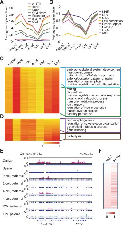
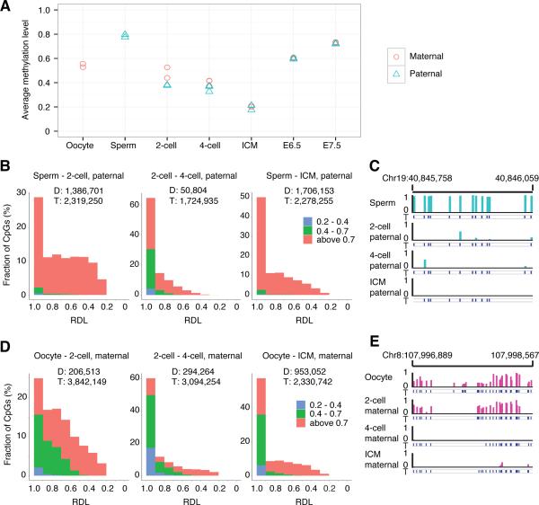
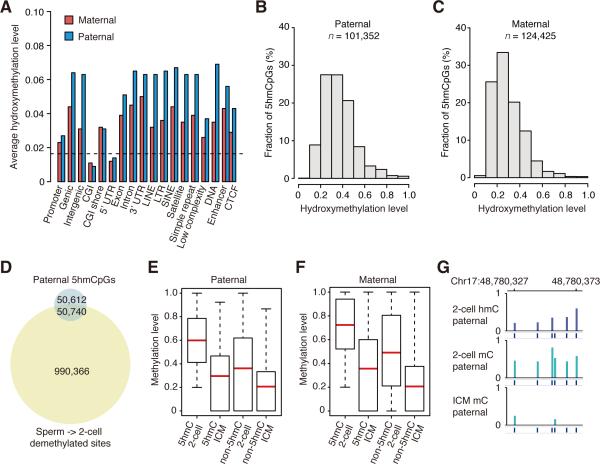
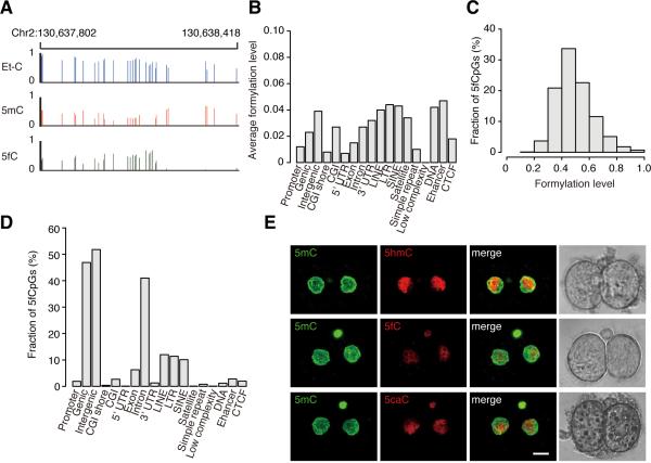
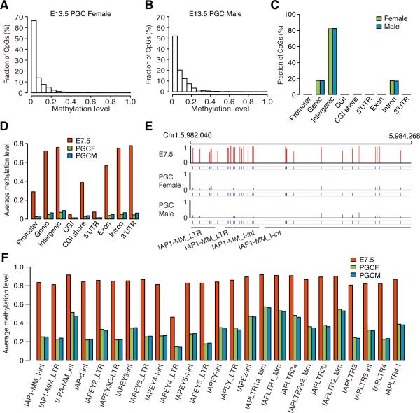
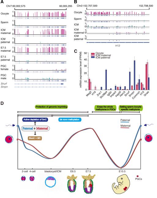
The reprogramming of parental methylomes is essential for embryonic development. In mammals, paternal 5-methylcytosines (5mCs) have been proposed to be actively converted to oxidized bases. These paternal oxidized bases and maternal 5mCs are believed to be passively diluted by cell divisions. By generating single-base resolution, allele-specific DNA methylomes from mouse gametes, early embryos, and primordial germ cell (PGC), as well as single-base-resolution maps of oxidized cytosine bases for early embryos, we report the existence of 5hmC and 5fC in both maternal and paternal genomes and find that 5mC or its oxidized derivatives, at the majority of demethylated CpGs, are converted to unmodified cytosines independent of passive dilution from gametes to four-cell embryos. Therefore, we conclude that paternal methylome and at least a significant proportion of maternal methylome go through active demethylation during embryonic development. Additionally, all the known imprinting control regions (ICRs) were classified into germ-line or somatic ICRs.


Figures

Figure 1
Figure 1. Dynamics of DNA methylomes for gametes and early embryos
(A) The dynamics of methylation levels of different genomic elements (genic related classification) during early embryogenesis. The average methylation is the mean value of the methylation levels of all CpGs located in the specific element. (B) The dynamics of methylation levels of different repeat elements. IAP is the sub-category of LTR. (C) and (D) Heat map of the methylation reprogramming of oocyte-specific hypomethylated promoters and sperm-specific hypo-methylated promoters during early embryogenesis respectively. GO term enrichment in genes with E7.5 hypomethylated promoters or hyper methylated promoters. DNA methylation level is colored from orange to red to indicate low to high. (E) Graphical representation of a genomic region showing the methylation level of non-CpGs for tracked maternal DNA (pink) and paternal DNA (light Blue) separately. Tracked non-CG cytosines are highlighted by short blue lines. “pa” means paternal DNA; “ma” means maternal DNA. (F) Positive-correlations between gene expression and methylated non-CG cytosines in the genic regions. Pearson correlation coefficiency is 0.20 (p value < 10−5). Each row represents one gene from the total 22,742 expressed genes. mC/C means the density of methylated non-CG cytosines. FPKM represents gene expression level. Both DNA methylation density and gene expression are colored from white to red to indicate low to high. See also Figure S1 and S2, Table S1, S2 and S3.
Figure 2
Figure 2. Both paternal and maternal DNAs are actively demethylated
(A) The dynamics of the average methylation level of paternal and maternal genome during early embryogenesis, respectively. (B) Distribution of paternal demethylated CpGs according to the RDL between two compared stages. Y axis represents the fraction of demethylated CpGs. X axis represents the relative demethylation level. “D” means the number of the demethylated CpGs between two compared stages; “T” means the number of CpGs covered in both two compared stages with at least 5 reads. The CpGs were categorized into three classes according to the methylation level indicated by different colors. For example, in the left panel, red indicates the highly methylated CpGs in sperm (methylation level (ML) >0.7), green indicates intermediate methylated CpGs in sperm (0.7≥ML>0.4), and blue indicates low methylated CpGs in sperm (0.4≥ML>0.2). 63% demethylated CpGs with RDL higher than 0.6 from sperm to 2-cell embryos; 95% demethylated CpGs with RDL higher than 0.6 from 2-cell embryos to 4-cell embryos; 82% demethylated CpGs with RDL higher than 0.6 from sperm to ICM. (C) Graphical representation of methylation pattern in paternal DNA at a locus in sperm, 2-cell embryos, 4-cell embryos, and ICM. The tracked CpGs are highlighted by short blue lines. (D) Distribution of maternal demethylated CpGs according to the RDL between two compared stages. 74% demethylated CpGs with RDLs higher than 0.6 from oocyte to 2-cell embryos; 86% demethylated CpGs with RDL higher than 0.6 from 2-cell embryos to 4-cell embryos; 84% demethylated CpGs with RDL higher than 0.6 from oocyte to ICM. (E) Graphical representation of methylation pattern from maternal DNA at a locus for oocyte, 2-cell, 4-cell, and ICM. See also Figure S3 and S4 and Table S4
Figure 3
Figure 3. 5hmC in both maternal and paternal genomes
(A) Average hydroxymethylation level of different genomic elements. The black dash line indicates the genomic background level (0.016). The average hydroxymethylation level is the mean value of the hydroxymethylation levels of all CpGs located in the specific element. Since there is no enhancer, CTCF data available for mouse 2-cell embryos, we used the data of mESC as the reference (Shen et al., 2012). (B) Distribution of 5hmCpGs in paternal genome according to the hydroxymethylation level. Y axis represents the fraction of 5hmCpGs. X axis represents the hydroxymethylation level. (C) Distribution of 5hmCpGs in maternal genome according to the hydroxymethylation level. (D) Venn diagram shows that a small proportion of CpGs are overlapped between 5hmCpGs in 2-cell embryos and paternal demethylated CpGs from sperm to 2-cell embryos. (E) Box plots of methylation levels of 5hmCpG sites or non-5hmCpG sites in paternal tracked DNA in 2-cell embryos and ICM. “5hmC” means 5hmCpGs. “non-5hmC” means non-5hmCpGs. Red line indicates the average methylation level, edges stand for the 25th/75th percentile, and whiskers stand for the 2.5th/97.5th percentile. (F) Box plots of methylation levels of 5hmCpG sites or non-5hmCpG sites in maternal tracked DNA in 2-cell embryos and ICM. (G) Graphical representation of 5hmC, 5mC pattern at a locus with paternal tracked CpGs in 2-cell embryos and ICM. See also Figure S5 and Table S5.
Figure 4
Figure 4. 5fC and 5caC in 2-cell embryos
(A) Graphical representation of the Et-CpG level, methylation level and formylation level at a locus in 2-cell embryos. (B) The average formylation level of different genomic elements in genome-wide DNA. The average formylation level is the mean value of the formylation levels of all CpGs located in the specific element. (C) Distribution of significant enriched 5fCpG sites according to the formylation level. Y axis represents the fraction of 5fCpGs. X axis represents the formylation level. (D) Distribution of the 5fCpG sites in the different genomic elements in 2-cell embryos. Y axis represents the fraction of 5fCpGs map to each genomic element versus all 5fCpGs. (E) Comparing the relative amount of 5hmC, 5fC and 5caC in 2-cell embryos with immunostaining. Antibody dilution is 1:2000 for Ab-5fC, 1:4000 for Ab-5hmC, and 1:5000 for Ab- 5caC. Scale bar is 10μm. See also Figure S6 and Table S6.
Figure 5
Figure 5. DNA methylation pattern for E13.5 PGCs
(A) Distribution of CpGs according to the methylation level in female E13.5 PGCs. (B) Distribution of CpGs according to the methylation level in male E13.5 PGCs. (C) The distribution of highly methylated CpGs (methylation level ≥ 0.8) in different genomic elements (genic related classification) for PGCs. (D) Average methylation levels of different genomic elements (genic related classification) in E7.5, male PGCs and female PGCs. (E) Graphical representation of methylation pattern of four IAPs in E7.5 embryos, male E13.5 PGCs, and female E13.5 PGCs. (F) Average methylation levels of all the different IAPs in E7.5 embryos, female E13.5 PGCs, and male E13.5 PGCs. See also Figure S7.
Figure 6
Figure 6. gICRs dynamics and imprinted gene expression in gametes and early embryos
(A) Graphical representation of the methylation pattern of gICR Snurf/Snrpn in gametes, ICM, E7.5 embryos and PGCs. The graph shows that the ICR is maintained in early embryos, but erased in E13.5 PGCs. (B) Graphical representation of the methylation pattern for gICR H13 DMR2 (3’ end) in gametes and ICM. The graph shows that this gICR is erased during early embryogenesis. (C) Allele-specific gene expression of previously defined imprinted genes in oocyte and ICM. Data are represented as mean +/- SEM of three independent repeats. (D) Two waves of genome-wide demethylation in mammals. 5hmC and 5fC present in both maternal and paternal genomes. Genome-wide demethylation is mainly through active demethylation in both paternal and maternal genomes, which is independent of the passive dilution of 5mC or its oxidized derivatives. See also Table 1 and Table S7.

References

    1. Collado-Fernandez E, Picton HM, Dumollard R. Metabolism throughout follicle and oocyte development in mammals. The International journal of developmental biology. 2012;56:799–808. - PubMed
    1. Feng S, Cokus SJ, Zhang X, Chen PY, Bostick M, Goll MG, Hetzel J, Jain J, Strauss SH, Halpern ME, et al. Conservation and divergence of methylation patterning in plants and animals. Proceedings of the National Academy of Sciences of the United States of America. 2010;107:8689–8694. - PMC - PubMed
    1. Ficz G, Branco MR, Seisenberger S, Santos F, Krueger F, Hore TA, Marques CJ, Andrews S, Reik W. Dynamic regulation of 5-hydroxymethylcytosine in mouse ES cells and during differentiation. Nature. 2011;473:398–402. - PubMed
    1. Gu TP, Guo F, Yang H, Wu HP, Xu GF, Liu W, Xie ZG, Shi L, He X, Jin SG, et al. The role of Tet3 DNA dioxygenase in epigenetic reprogramming by oocytes. Nature. 2011;477:606–610. - PubMed
    1. Hackett JA, Sengupta R, Zylicz JJ, Murakami K, Lee C, Down TA, Surani MA. Germline DNA Demethylation Dynamics and Imprint Erasure Through 5-Hydroxymethylcytosine. Science. 2013;339:448–452. - PMC - PubMed

Publication types

Associated data