. 2014 Aug;9(8):648-655.
doi: 10.1038/nnano.2014.84. Epub 2014 May 11.

In vivo endothelial siRNA delivery using polymeric nanoparticles with low molecular weight

Affiliations

In vivo endothelial siRNA delivery using polymeric nanoparticles with low molecular weight

James E Dahlman et al. Nat Nanotechnol. 2014 Aug.

Abstract

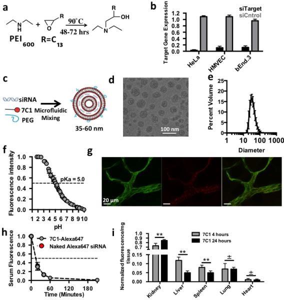
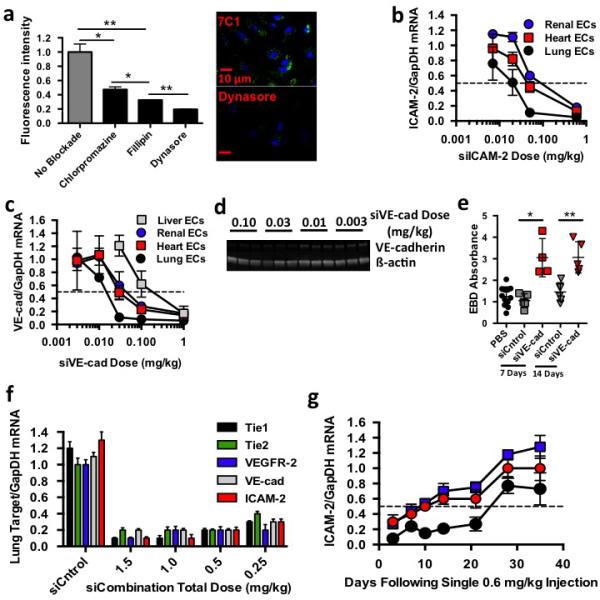
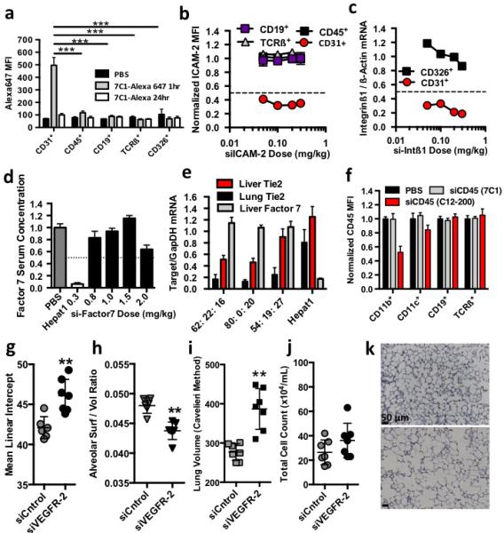
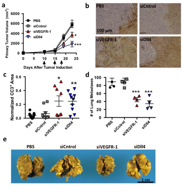
Dysfunctional endothelium contributes to more diseases than any other tissue in the body. Small interfering RNAs (siRNAs) can help in the study and treatment of endothelial cells in vivo by durably silencing multiple genes simultaneously, but efficient siRNA delivery has so far remained challenging. Here, we show that polymeric nanoparticles made of low-molecular-weight polyamines and lipids can deliver siRNA to endothelial cells with high efficiency, thereby facilitating the simultaneous silencing of multiple endothelial genes in vivo. Unlike lipid or lipid-like nanoparticles, this formulation does not significantly reduce gene expression in hepatocytes or immune cells even at the dosage necessary for endothelial gene silencing. These nanoparticles mediate the most durable non-liver silencing reported so far and facilitate the delivery of siRNAs that modify endothelial function in mouse models of vascular permeability, emphysema, primary tumour growth and metastasis.


Figures

Figure 1
Figure 1
7C1 synthesis, characteri zation, and in vivo biodistribution. (A) 7C1 synthesis scheme. (B) Target gene expression 24 hours following 30 nM treatment with siRNA in human cervical carcinoma (HeLa), human primary endothelial (HMVEC), and murine endothelial (bEnd.3) cells. HeLa target gene expression was measured as Firefly luminescence in HeLa cells expressing Luciferase that were treated with siRNA targeting luciferase. bEnd.3 and HMVEC target gene expression was measured as Tie2 mRNA levels following treatment with siRNA targeting Tie2. (C) 7C1 formulation scheme. 7C1 nanoparticles were mixed with C14PEG2000 and siRNA in a high throughput microfluidic chamber as previously described. (D) 7C1 internal structure characterized by cryo-TEM. Dark bands indicate lipid layers and light bands indicate regions with siRNA. (E) Average 7C1 hydrodynamic diameter, measured by dynamic light scattering, and weighted by volume (N=20 formulations). (F) TNS nuorescence of formulated 7C1 nanoparticles as a function of pH (used to measure 7C1 pKa). (G) Representative confocal image of Alexa647-tagged siRNA complexed to 7C1 one hour after intravenous injection. CD31 is a ubiquitous marker for endothelium (Scale bar = 20 μm). (H) Serum Cy5.5 concentration following with 7C1-Cy5.5 siRNA or naked Cy5.5 siRNA (I) Cy5.5 nuorescence/mg tissue after injection with 7C1-Cy5.5 siRNA. Tissues were removed after injection and weighed individually. Cy5.5 intensity was normalized to each individual tissue. Timepoints were selected to measure systemic siRNA accumulation after Cy5.5 was cleared from serum. N=4-S mice/group. In all cases, data shown as mean +/− std. *p<0.05, ** p<0.005, ***p<:0.0008, ±p<0.75.
Figure 2
Figure 2
7Cl delivers siRNA to endothelial cells. (A) Alexa647 fluorescence uptake in HMVEC cells following 7CI-Alexa647 siRNA treatments and administration of small molecules blockading clathrin (Chlorpromazine), caveolin (Fillipin), and both endocytotic pathways (Oynasore). (B) ICAM-2/GapDH mRNA ratios (normalized to PBS-treated mice) following intravenous injection of 7Cl-silCAM-2. (C) VE-cadherin/GapDH mRNA ratios (normalized to PBS-treated mice) following intravenous injection of 7Cl-siVEcad. (D) VE-cadherin and ß-actin protein expression following treatment with 7Cl-siVEcad. (E) Evans Blue Dye pulmonary absorbance seven and fourteen days following a 0.6 mg/kg injection of 7Cl-siVEcad. (F) Target/GapDH mRNA ratios (normalized to PBS-treated mice) following injection of 7Cl formulated with siCntrol or five siRNAs targeting ICAM-2, Tie2, VE-cadherin, VEGFR2, or Tiel, respectively (siCombination). (G) ICAM-2/GapDH mRNA levels as a function of time following a 0.6 mg/kg injection of siICAM-2. Data shown as mean +/− std. N=4 to 5 mice per group, *p< 0.05, ** p< 0.005.
Figure 3
Figure 3
7Cl preferentially delivers siRNA to pulmonary endothelial cells in vivo. (A) Alexa647 median fluorescent intensity in pulmonary endothelial (C031+), hematopoietic (C045+), epithelial (C0326+), B (CDI9+), or T (TCRß+) cells isolated from mice after treatment with 7Cl formulated with Alexa647-tagged siRNA. Statistical significance calculated between endothelial cells and other pulmonary cell types one hour after injection. (B) ICAM-2 median fluorescent intensity in pulmonary cells (normalized to siCntrol-treated mice) isolated from mice three days followings treatment with 7Cl-silCAM -2. (C) Integrinß/ß-actin mRNA ratios (normalized to siCntrol-treated mice) in pulmonary endothelial and epithelial cells isolated from mice two days after treatment with silntegrinßl. (D) Factor 7 serum concentration (normalized to PBS-treated animals) two days following treatment with liver targeting molecule HepatOl-siFactor7 or 7Cl-siFactor7 (E) Tie2 and Factor7/GapOH mRNA expression following a 0.15 mg/kg injection of 7Cl concurrently formulated with siTie2 and siFactor7. Particles were formulated with different 7Cl: Cholesterol: C14PEGlOOO molar ratios. 7Cl decreased Tie2 mRNA expression in pulmonary, renal, and hepatic endothelium without reducing F7 mRNA expression. (F) C045 median fluorescent intensity following treatment with 7Cl-siC04S or positive control CI2-200-siC045. (G-K) Mean Linear Intercept (MU) between alveoli, pulmonary surface/volume ratio, total volume, and pulmonary histology following two 0.5 mg/kg injections of siCntrol or siVEGFR2. Increased MLI, alveolar volume, decreased surface/volume ratios, and constant infiltrating myeloid cells are consistent with an induced emphysema-like phenotype (N=6 to 7 animals / group, data shown as average +/− std, scale bar = 50 um)*p < 0.05, **p<0.002, ***p<0.00l.
Figure 4
Figure 4
7Cl mediated mRNA silencing modifies endothelial function in vivo. (A) Primary lewis lung Carcinoma (LLC) growth following three 1.0 mg/kg treatments with PBS, siCntrol. siVEGFR-l, or siDII4 (N=7 to 10 animals per group, data shown as average +/− SEM). (B,C) Representative image and quantification of cleaved caspase 3 (CC3) staining, a marker for apoptosis, following treatment with PBS, siCntrol, siVEGFR-l, siDlI4. Normalized CC3+ area defined as the total CC3+ surface area divided by the tumor surface area. Scale bar = 100 μm. (D) Number of pulmonary surface metastases following four 1.0 mg/kg injections with PBS, siCntrol. siVEGFR-l, or siDli4 (N=4 to 6 per group, data shown as average +/− SEM). To measure effects independent of primary tumor growth, animals were not treated until after primary tumor resection. (E) Murine lungs with metastatic lesion removed after treatment with PBS, siluc, siVEGFR-l, or siDlI4 . *p < 0.05, **p<0.002, ***p<0.00l.

Comment in

References

    1. Pober JS, Sessa WC. Evolving functions of endothelial cells in inflammation. Nat Rev Immunol. 2007;7:803–815. - PubMed
    1. Hagberg CE, et al. Targeting VEGF-B as a novel treatment for insulin resistance and type 2 diabetes. Nature. 2012;490:426–430. - PubMed
    1. Kumar V, Abbas A, Fausto N, Aster J. Robbins and Cotran Pathologic Basis of Disease. (Eighth Edition) 2009
    1. Kanasty R, Dorkin JR, Vegas A, Anderson D. Delivery materials for siRNA therapeutics. Nat Mater. 2013;12:967–977. - PubMed
    1. Love KT, et al. Lipid-like materials for low-dose, in vivo gene silencing. Proc Natl Acad Sci USA. 2010;107:1864–1869. - PMC - PubMed

Publication types