. 2014 May 22;7(4):1320-32.
doi: 10.1016/j.celrep.2014.04.002. Epub 2014 May 9.

Isolation of chromatin from dysfunctional telomeres reveals an important role for Ring1b in NHEJ-mediated chromosome fusions

Affiliations

Isolation of chromatin from dysfunctional telomeres reveals an important role for Ring1b in NHEJ-mediated chromosome fusions

Cristina Bartocci et al. Cell Rep. .

Abstract

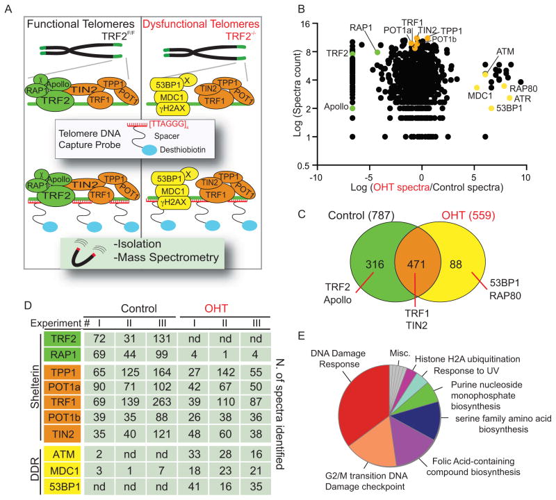
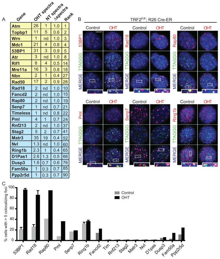
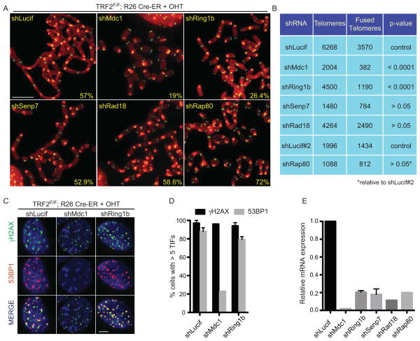
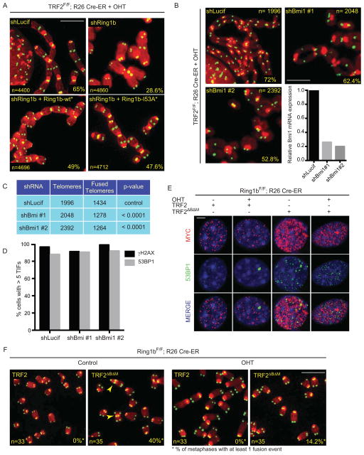
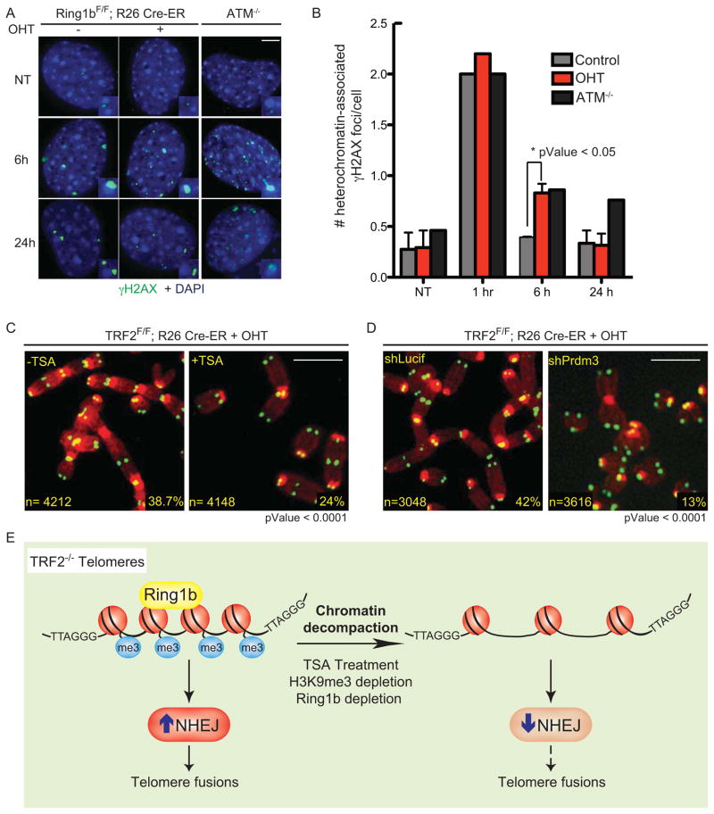
When telomeres become critically short, DNA damage response factors are recruited at chromosome ends, initiating a cellular response to DNA damage. We performed proteomic isolation of chromatin fragments (PICh) in order to define changes in chromatin composition that occur upon onset of acute telomere dysfunction triggered by depletion of the telomere-associated factor TRF2. This unbiased purification of telomere-associated proteins in functional or dysfunctional conditions revealed the dynamic changes in chromatin composition that take place at telomeres upon DNA damage induction. On the basis of our results, we describe a critical role for the polycomb group protein Ring1b in nonhomologous end-joining (NHEJ)-mediated end-to-end chromosome fusions. We show that cells with reduced levels of Ring1b have a reduced ability to repair uncapped telomeric chromatin. Our data represent an unbiased isolation of chromatin undergoing DNA damage and are a valuable resource to map the changes in chromatin composition in response to DNA damage activation.


Figures

Figure 1
Figure 1. Proteomics of isolated chromatin segments (PICh) of functional and dysfunctional mouse telomeres
A. Schematic representation of the PICh analysis employed to define the chromatin changes that occur at telomeres upon TRF2 depletion. B. Identified proteins were plotted based on the Log2 Intensity (Y axis) calculated based on the total spectra counts identified and ratio between TRF2 proficient and deficient settings (X axis) calculated as Log2 (OHT spectra/control spectra). Only proteins identified in at least 2 experiments are plotted. Green and orange dots represent proteins recruited at telomeres in a TRF2-dependent or independent manner, respectively. Yellow dots represent known DNA damage factors recruited to dysfunctional telomeres. C. Venn diagrams depicting the total number of proteins identified in at least 2 out of 3 PICh experiments from the control and OHT samples and the degree of overlap between the datasets from the two samples. D. Table listing shelterin components and some of the DNA damage response (DDR) factors identified with the corresponding spectra counts isolated in each individual experiment (nd= not detected). E. Pie chart illustrating the distribution of proteins identified in the TRF2-depleted samples (OHT), classified according to Gene Ontology (GO). Note the enrichment for DNA damage response proteins
Figure 2
Figure 2. Validation of novel factors that localize at dysfunctional telomeres
A. List of proteins identified by PICh as enriched at dysfunctional telomeres. Average number of spectra identified at TRF2 deficient (OHT) or proficient (NT) telomeres is indicated. Proteins were ranked based on their relative enrichment at TRF2 depleted telomeres and normalized to absolute expression of the protein in mouse cells (PaxDB database). Proteins highlighted in yellow represent DNA damage response factors known to localize at dysfunctional telomeres. Proteins highlighted in blue represent proteins selected for validation. B. Localization of ectopically expressed tagged proteins (myc/GFP) (stained in red) to telomere repeats (TTAGGG, stained in green). TRF2F/F Rosa26 CRE-ER MEFs infected with the indicated constructs were treated with OHT and harvested 3 days later. Scale bar 5 μm. C. Quantification of data shown in B: cells with 5 or more foci co-localizing with telomeres were counted as positive. Error bars represent standard deviation (SD) of average of three independent experiments. See also Figure S2.
Figure 3
Figure 3. Ring1b plays a critical role in the response to dysfunctional telomeres
A. Metaphase spreads were harvested from TRF2F/F Rosa26 CRE-ER MEFs infected with a control lentiviral construct (shLucif) or with the indicated shRNA constructs and treated with OHT for 96 hours. Chromosomes were stained with a PNA probe complementary to telomeric repeats (green) and DAPI (red). Percentages of fused chromosome ends are indicated. Scale bar 5 μm. B. Quantification of data shown in panel A. C. TRF2F/F Rosa26 CRE-ER MEFs infected with the indicated shRNA constructs were treated with OHT for 72 hours, fixed and stained for γH2AX (green), 53BP1 (red) and DAPI (blue). Scale bar 5 μm. D. Quantification of the data shown in C (cells with 5 or more γH2AX/53BP1 foci were counted as positive). Error bars represent SD of average of three independent experiments. E. Quantification of relative mRNA levels of the indicated genes in cells infected with the indicated shRNA constructs, normalized to shLucif as control. See also Figure S3.
Figure 4
Figure 4. The Ring1b/Bmi1 heterodimer acts on the chromatin at dysfunctional telomeres
A. Metaphase spreads of TRF2F/F Rosa26 CRE-ER MEFs infected with shLuciferase or shRing1b and Ring1b-wt* or Ring1b-I53A* and treated with OHT for 96 hours. Percentages of fused chromosome ends are indicated. Scale bar 5 μm. B. Metaphase spreads of TRF2F/F Rosa26 CRE-ER MEFs infected with shRNA constructs directed against Bmi1 and treated with OHT for 96 hours. Percentages of fused chromosome ends are indicated. Scale bar 5 μm. Graph shows quantification of relative Bmi1 mRNA levels measured by qPCR, normalized to shLucif as control. C. Quantification of end-to-end chromosome fusions in B. D. Quantification of γH2AX- and 53BP1-positive TRF2F/F Rosa26 CRE-ER MEFs infected as described in B, treated with OHT for 72 hours, fixed and stained for γH2AX and 53BP1 (cells with 5 or more γH2AX/53BP1 were counted as positive). E. Ring1bF/F Rosa26 CRE-ER MEFs (−/+ OHT, 96 hours) were infected with wild type TRF2 or a TRF2 dominant negative allele (TRF2ΔBΔM) as indicated, fixed and stained for myc (red), 53BP1 (green) and DAPI (blue). Scale bar 5 μm. F. Metaphase spreads of Ring1bF/F Rosa26 CRE-ER MEFs treated as in E were stained for telomeric DNA (Green) to detect end-to-end chromosome fusions (arrowheads). Number of metaphases analyzed and percentage of metaphases with at least 1 fusion event are indicated. Scale bar 5 μm. See also Figure S4.
Figure 5
Figure 5. Compact chromatin favors NHEJ-mediated repair of dysfunctional telomeres
A. Confluent, stationary-phase Ring1bF/F Rosa26 CRE-ER MEFs untreated or treated with tamoxifen (−/+ OHT) were left untreated (NT) or irradiated (2 Gy) and harvested at the indicated time points post irradiation. ATM−/− MEFs were used as control. Cells were fixed and stained for γH2AX (green) and DAPI (blue). Scale bar 5 μm. B. Quantification of the total number of heterochromatin-associated γH2AX foci in cells treated as described in A. Error bars represent SD of average of three independent experiments. C. Metaphase spreads of TRF2F/F Rosa26 CRE-ER MEFs treated with OHT for 72 hours and when indicated with TSA (18 hours). Percentages of fused chromosome ends are indicated. Scale bar 5 μm. D. Metaphase spreads of TRF2F/F Rosa26 CRE-ER MEFs infected with a shRNA against Prdm3 (shPrdm3) or a control shRNA (shLucif) and treated with OHT for 72 hours. Scale bar 5 μm. E. Model for the role of chromatin compaction in NHEJ-mediated repair of dysfunctional telomeres. See also Figure S5.

References

    1. Artandi SE, Chang S, Lee SL, Alson S, Gottlieb GJ, Chin L, DePinho RA. Telomere dysfunction promotes non-reciprocal translocations and epithelial cancers in mice. Nature. 2000;406:641–645. - PubMed
    1. Attwooll CL, Akpinar M, Petrini JH. The mre11 complex and the response to dysfunctional telomeres. Molecular and cellular biology. 2009;29:5540–5551. - PMC - PubMed
    1. Augereau A, T’Kint de Roodenbeke C, Simonet T, Bauwens S, Horard B, Callanan M, Leroux D, Jallades L, Salles G, Gilson E, et al. Telomeric damage in early stage of chronic lymphocytic leukemia correlates with shelterin dysregulation. Blood. 2011;118:1316–1322. - PubMed
    1. Bannister AJ, Zegerman P, Partridge JF, Miska EA, Thomas JO, Allshire RC, Kouzarides T. Selective recognition of methylated lysine 9 on histone H3 by the HP1 chromo domain. Nature. 2001;410:120–124. - PubMed
    1. Buchwald G, van der Stoop P, Weichenrieder O, Perrakis A, van Lohuizen M, Sixma TK. Structure and E3-ligase activity of the Ring-Ring complex of polycomb proteins Bmi1 and Ring1b. The EMBO journal. 2006;25:2465–2474. - PMC - PubMed

Publication types