. 2014 Jun 19;510(7505):422-426.
doi: 10.1038/nature13263. Epub 2014 May 11.

Ribosomal oxygenases are structurally conserved from prokaryotes to humans

Affiliations

Ribosomal oxygenases are structurally conserved from prokaryotes to humans

Rasheduzzaman Chowdhury et al. Nature. .

Abstract

2-Oxoglutarate (2OG)-dependent oxygenases have important roles in the regulation of gene expression via demethylation of N-methylated chromatin components and in the hydroxylation of transcription factors and splicing factor proteins. Recently, 2OG-dependent oxygenases that catalyse hydroxylation of transfer RNA and ribosomal proteins have been shown to be important in translation relating to cellular growth, TH17-cell differentiation and translational accuracy. The finding that ribosomal oxygenases (ROXs) occur in organisms ranging from prokaryotes to humans raises questions as to their structural and evolutionary relationships. In Escherichia coli, YcfD catalyses arginine hydroxylation in the ribosomal protein L16; in humans, MYC-induced nuclear antigen (MINA53; also known as MINA) and nucleolar protein 66 (NO66) catalyse histidine hydroxylation in the ribosomal proteins RPL27A and RPL8, respectively. The functional assignments of ROXs open therapeutic possibilities via either ROX inhibition or targeting of differentially modified ribosomes. Despite differences in the residue and protein selectivities of prokaryotic and eukaryotic ROXs, comparison of the crystal structures of E. coli YcfD and Rhodothermus marinus YcfD with those of human MINA53 and NO66 reveals highly conserved folds and novel dimerization modes defining a new structural subfamily of 2OG-dependent oxygenases. ROX structures with and without their substrates support their functional assignments as hydroxylases but not demethylases, and reveal how the subfamily has evolved to catalyse the hydroxylation of different residue side chains of ribosomal proteins. Comparison of ROX crystal structures with those of other JmjC-domain-containing hydroxylases, including the hypoxia-inducible factor asparaginyl hydroxylase FIH and histone N(ε)-methyl lysine demethylases, identifies branch points in 2OG-dependent oxygenase evolution and distinguishes between JmjC-containing hydroxylases and demethylases catalysing modifications of translational and transcriptional machinery. The structures reveal that new protein hydroxylation activities can evolve by changing the coordination position from which the iron-bound substrate-oxidizing species reacts. This coordination flexibility has probably contributed to the evolution of the wide range of reactions catalysed by oxygenases.


Figures

Figure 1
Figure 1. The overall folds of the ribosomal oxygenases
a, Reactions catalyzed by ROX and related oxygenases. CAD: C-terminal transactivation domain of HIF-α; ARD: Ankyrin repeat domain. b, Ribbons representations of ycfD, ycfDRM, Mina53 and NO66 homodimers. The monomers contain a JmjC domain with the double-stranded-β-helix (DSBH) core present in all 2OG-oxygenases (blue) followed by dimerization (yellow) and C-terminal ‘winged-helix’ domains (red). Domain architecture and a schematic representation of the DSBH core β-strands (βI-VIII) that form major (grey, βI, VIII, III and VI) and minor sheets (blue, βII, VII, IV and V) is shown boxed. The insert between βIV and βV (purple) is involved in substrate binding. The 3 Fe-coordinating residues are on the βII and βVII strands (black circles). 2OG is in green sticks; the 2OG C5-carboxylate binding residue, Arg (ycfDs) or Lys (hROX) from βIV is a black circle.
Figure 2
Figure 2. Comparison of the substrate structures for ROX/JmjC enzymes
Ribbons representations from ROX and related 2OG-oxygenase-substrate complexes: a, Mina53·Mn·2OG·rpL27a(32-50) (P212121, 2.05 Å), b, NO66·Mn·NOG·rpL8(205-224) (C2, 2.35 Å), c, ycfDRM·Mn·NOG·L16(72-91) (P212121, 3.0 Å), d, FIH·Fe·NOG·HIF-1α(786-826) (PDB: 1H2K), e, PHF8·Fe·NOG·histone H3K4me3K9me2(2-25) (PDB: 3KV4), f, KDM4A·Ni·NOG·histone H3K9me2(7-14) (PDB: 2OX0). For comparison, the DSBH core of each structure is in a similar orientation. Note the directionality of substrate binding in the JmjC domains. The active site metals (Fe/ surrogate) are color-coded spheres. Analyses of the structures reveal that the ROX overall folds (a-c), oligomerization states and active site architectures are evolutionarily conserved.
Figure 3
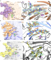
Figure 3. Features of ROX-substrate binding
Ribbons representations of Mina53 (a), NO66 (b) and ycfDRM (c) monomers showing difference electron density (Fo-Fc OMIT) for substrates contoured to 3σ (right panels). Left panels show active site surface representations, showing key hydrogen-bonds/polar interactions (dotted lines) with substrates. a, With Mina53, the His39rpL27a imidazole nitrogens form hydrogen-bonds with Tyr167/Ser257 (NδHis39-OHTyr167 2.9 Å; NεHis39-OγSer257 3.1 Å). b, In NO66, His216rpL8 is similarly bound in a deep pocket; the His216rpL8 imidazole nitrogens form hydrogen-bonds with Tyr328/Ser421 (NδHis216-OHTyr328 3.2 Å; NεHis216-OγSer421 2.7 Å) and hydrophobic interactions with Ile244, that project its pro-S hydrogen toward the metal (metal-β-CH2, 4.4 Å). While Mina53 (a) uses 4 primary amides, Asn101, Gln136, Gln139 and Asn165 to interact with rpL27a backbone amides, NO66 (b) uses 2 arginines (272, 297) to hydrogen-bond with the Asn215rpL8 sidechain and His216rpL8 backbone. In the ycfDRM·L16 complex (c), Arg82L16 binds in a pocket defined by the Tyr137/Met120 sidechains, which form π-cation and hydrophobic interactions with Arg82L16 sidechain. The Arg82 guanidino group makes electrostatic interactions with the Asp118ycfDRM carboxylate (O-NH, 2.8-3.1 Å) and hydrogen-bonds to Ser208ycfDRM (NεArg82-OHSer208 3.5 Å; NηArg82-COSer208 3.2 Å). Although Tyr167Mina53/Tyr328NO66 are not positionally related to Tyr137ycfDRM, the role of the serine (Ser257Mina53, Ser421NO66, Ser208ycfDRM, β-VIII) in binding the hydroxylated His/Arg is conserved in ROX. Substitutions of these residues cause significant loss of activity (see Extended Data Fig. 6).
Figure 4
Figure 4. Proposed sequence of evolution of active metal chemistry of ROX and related JmjC 2OG-oxygenases
The figure compares views from active sites of representative JmjC-enzymes and suggests how the ROX fold evolved into JmjC-hydroxylases and -KDMs. Structurally informed cross-genomic bioinformatic analyses imply that the ROX are the earliest identified JmjC 2OG-oxygenases; ycfD and NO66 both exist in prokaryotes but only NO66 is identified in eukaryotes. Coupled to the analyses of the active sites, these analyses imply NO66/close-relatives are the precursors of Mina53 and other JmjC-hydroxyalses and KDMs. a, Upper panel: structure based alignment of ROX, FIH, PHF8 and KDM4A with DSBH core labeled βI-VIII, iron-coordinating and the 2OG C5-carboxylate binding residues in red and green. Lower panels: analyses of active sites suggest conservation of metal-/2OG-binding in ROX, FIH and KDMs, note the 2OG C5-carboxylate binding residue (usually from βIV in JmjC-enzymes), changes from an Arg (in ycfDs) to a Lys (in hROX, JmjC-hydroxyalses/KDMs) (Extended Data Fig. 4). b, Overlays of the NO66/ycfDRM, NO66/Mina53, NO66/FIH, NO66/KDM4A active site views. The hydroxylated β-methylenes nearly superimpose in ROX, such that the oxidized C-H bonds (red arrows, 3-pro-R in Arg82L16 and 3-pro-S in His39rpL27a and His216rpL8) project toward the metal. The spatial relationship of the hydroxylated C3/Nε-methyl carbon with respect to the metal (and associated reactive oxidizing species) is conserved in ROX and the demethylases, e.g. KDM4A, but not in FIH. Note the different hydroxylation positions, but the similar orientation of Asn803CAD/FIH (hydroxylated) and Asn215rpL8/NO66 (not hydroxylated).

Similar articles

Cited by

References

    1. Klose RJ, Zhang Y. Regulation of histone methylation by demethylimination and demethylation. Nat. Rev. Mol. Cell. Biol. 2007;8:307–318. - PubMed
    1. Walport LJ, Hopkinson RJ, Schofield CJ. Mechanisms of human histone and nucleic acid demethylases. Curr. Opin. Chem. Biol. 2012;16:525–534. - PubMed
    1. Kaelin WG, Jr., Ratcliffe PJ. Oxygen sensing by metazoans: the central role of the HIF hydroxylase pathway. Mol. Cell. 2008;30:393–402. - PubMed
    1. Webby CJ, et al. Jmjd6 catalyses lysyl-hydroxylation of U2AF65, a protein associated with RNA splicing. Science. 2009;325:90–93. - PubMed
    1. Fu Y, et al. The AlkB domain of mammalian ABH8 catalyzes hydroxylation of 5-methoxycarbonylmethyluridine at the wobble position of tRNA. Angew. Chem. Int. Ed. Engl. 2010;49:8885–8888. - PMC - PubMed

Publication types