Review
. 2014 Jul;22(7):406-16.
doi: 10.1016/j.tim.2014.04.003. Epub 2014 May 7.

Clostridium difficile spore biology: sporulation, germination, and spore structural proteins

Affiliations
Review

Clostridium difficile spore biology: sporulation, germination, and spore structural proteins

Daniel Paredes-Sabja et al. Trends Microbiol. 2014 Jul.

Abstract

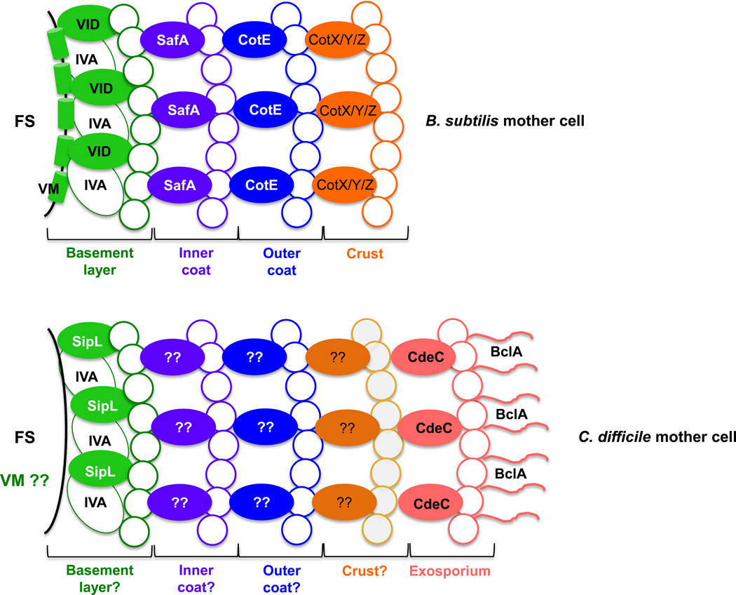
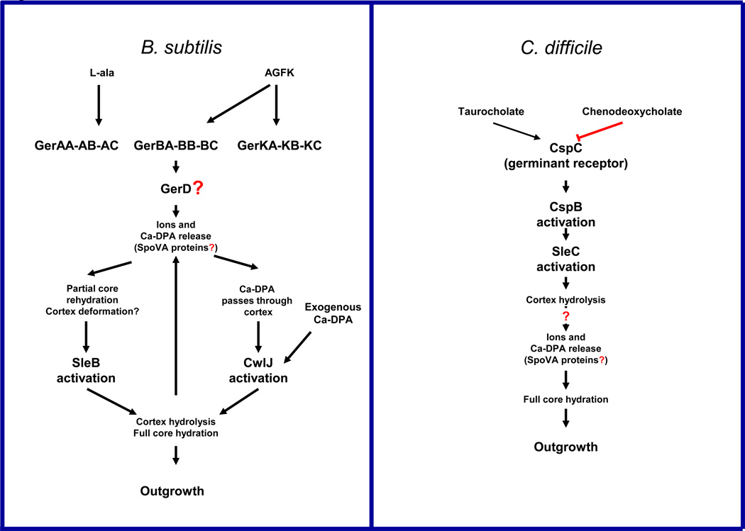
Clostridium difficile is a Gram-positive, spore-forming obligate anaerobe and a major nosocomial pathogen of worldwide concern. Owing to its strict anaerobic requirements, the infectious and transmissible morphotype is the dormant spore. In susceptible patients, C. difficile spores germinate in the colon to form the vegetative cells that initiate Clostridium difficile infections (CDI). During CDI, C. difficile induces a sporulation pathway that produces more spores; these spores are responsible for the persistence of C. difficile in patients and horizontal transmission between hospitalized patients. Although important to the C. difficile lifecycle, the C. difficile spore proteome is poorly conserved when compared to members of the Bacillus genus. Further, recent studies have revealed significant differences between C. difficile and Bacillus subtilis at the level of sporulation, germination, and spore coat and exosporium morphogenesis. In this review, the regulation of the sporulation and germination pathways and the morphogenesis of the spore coat and exosporium will be discussed.

Keywords: C. difficile spores; exosporium; germination; spore coat; sporulation.


Figures

Figure 1
Figure 1. Ultrastructure of Clostridium difficile spores
(A) Micrographs of thin sections of spores from various C. difficile strains. Upper and lower panels show TEM analyses of strains 630 ribotype 027 (RT12), R20291 RT27, M120 RT78, TL176 RT14, TL178 RT02. It is worth noting that some strains such as 630 and R20291 and TL178 have two different ultrastructural phenotypes. In the upper panel selected spores with a typical exosporium-like structure with roughed electro-dense material are shown, while in the lower panels representative micrographs of spores with less electron dense exosporium-like ultrastructures are shown, suggesting a deficient exosporium layer. Abbreviations: Ex, exosporium; Ct, coat; Cx, cortex. (B) For comparison, the ultrastructure of Bacillus anthracis spores is shown ([37], reprinted with permission of Wiley). Abbreviations: Ex-BL, exosporium basal-layer; Ex-N, Exosporium hair-like nap; Ct, coat; Cx, cortex. (C) The ultrastructure of the spore surface of representative spores of strains C. difficile TL176 and TL178 are shown, highlighting the presence of a highly organized surface of unknown exosporium proteins.
Figure 2
Figure 2. Main morphogenetic stages of the process of sporulation
Upon nutrient limitation, cells cease growth and initiate sporulation. Asymmetric cell division generates a smaller forespore compartment and a larger mother cell. Following completion of DNA segregation, the mother cell proceeds to engulf the forespore. During the initiation of spore metabolic dormancy and compaction of the spore DNA, the mother cell mediates the development of the forespore into the spore through the production of the cortex and the inner and outer coats. Next, the mother cell lyses and releases the mature spore. In the presence of the appropriate environmental stimulus, the dormant spore initiates the process of germination that leads to the re-initiation of vegetative growth. Abbreviations: MC, mother cell compartment; FS, forespore compartment; MS, mature spore.
Figure 3
Figure 3. Schematic of B. subtilis and C. difficile surface layers
The major proteinaceous layers defined by McKenney et al. [42] are shown. B. subtilis lacks an exosporium layer. Based on localization of SpoIVA and SpoVID in B. subtilis, C. difficile SpoIVA and SipL are shown localizing to a putative basement layer. It is unclear whether C. difficile SpoVM localizes to the forespore membrane similar to B. subtilis SpoVM (indicated by VM??) [42]. Morphogenetic proteins required for the assembly of the indicated layer are labeled; morphogenetic proteins for the putative inner and outer coat layers of C. difficile are unknown (? indicates that it remains unclear whether discrete layers defined by specific morphogenetic proteins as defined in B. subtilis exist in C. difficile spores). It is also unclear whether C. difficile spores have a crust layer. The morphogenetic protein of the exosporium layer, CdeC, and the outer most exosporium protein, BclA1, are shown. The location of BclA2 and BclA3 remain unknown. Abbreviations: FS, forespore compartment; VM, SpoVM; VID, SpoIVD; IVA, SpoIVA; ??, unidentified proteins.
Figure I Box I
Figure I Box I
Sporulation in endospore-forming B. subtilis and C. difficile. Models of the global regulatory process of the four main sporulation specific sigma factors in B. subtilis and C. difficile.
Figure I Box 2
Figure I Box 2
Structural layers of bacterial spores. The main layers of bacterial spore structure are shown and not drawn to scale. The exosporium and spore coat may have sub-layers which are not shown. The exosporium layer is present in some species. Figure has been modified from [75] with permission from Elsevier.
Figure I Box 3
Figure I Box 3
Tentative models for information flow during spore germination of B. subtilis and C. difficile. Arrows denote the preference of the germinants for the GRs, and upward arrows indicate that cortex hydrolysis might increase the rate of DPA release. Red line indicates that chenodeoxycholate inhibits CspC activation. Question marks indicate that there is suggestive, but no conclusive experimental evidence; AGFK, is a mixture of L-asparagine, glucose, fructose and KCl.

References

    1. Rupnik M, et al. Clostridium difficile infection: new developments in epidemiology and pathogenesis. Nat Rev Microbiol. 2009;7:526–536. - PubMed
    1. Kelly CP, LaMont JT. Clostridium difficile--more difficult than ever. N Engl J Med. 2008;359:1932–1940. - PubMed
    1. McFarland LV. Epidemiology, risk factors and treatments for antibiotic-associated diarrhea. Dig Dis. 1998;16:292–307. - PubMed
    1. Hernández-Rocha C, et al. Infecciones causadas por Clostridium difficile: una visión actualizada. Revista Chilena de Infectología. 2012;29:434–445. - PubMed
    1. Miller BA, et al. Comparison of the burdens of hospital-onset, healthcare facility-associated Clostridium difficile Infection and of healthcare-associated infection due to methicillin-resistant Staphylococcus aureus in community hospitals. Infect Control Hosp Epidemiol. 2011;32:387–390. - PubMed

Publication types

MeSH terms

Substances