Review
. 2014;90(5):163-83.
doi: 10.2183/pjab.90.163.

Functional zonation of the rat adrenal cortex: the development and maintenance

Affiliations
Review

Functional zonation of the rat adrenal cortex: the development and maintenance

Fumiko Mitani. Proc Jpn Acad Ser B Phys Biol Sci. 2014.

Abstract

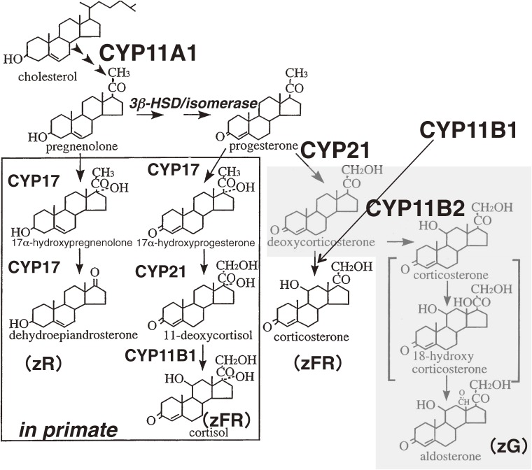
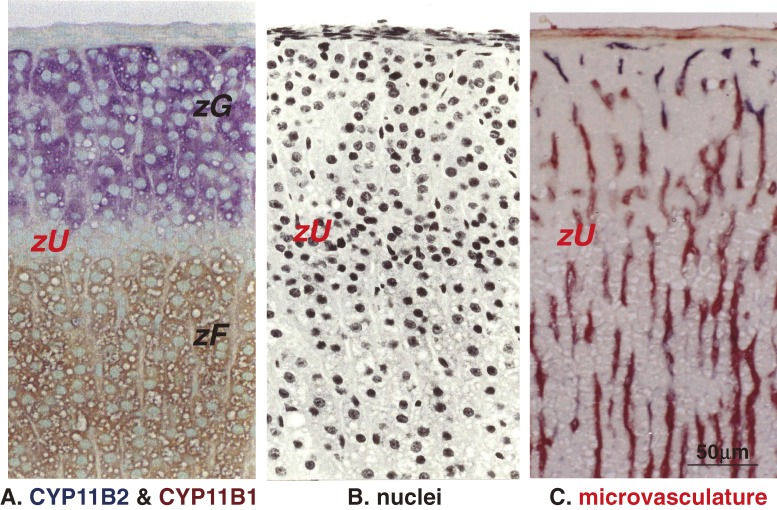
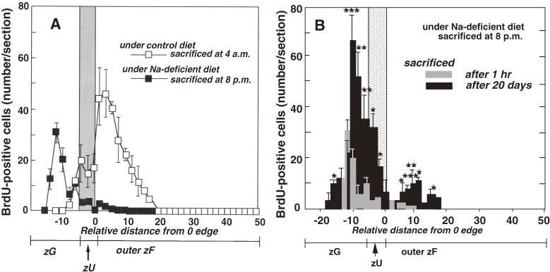
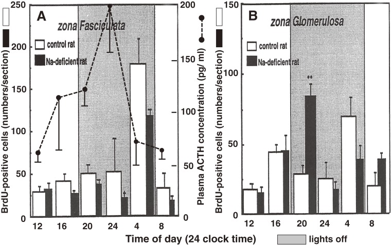
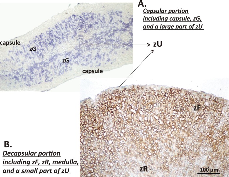
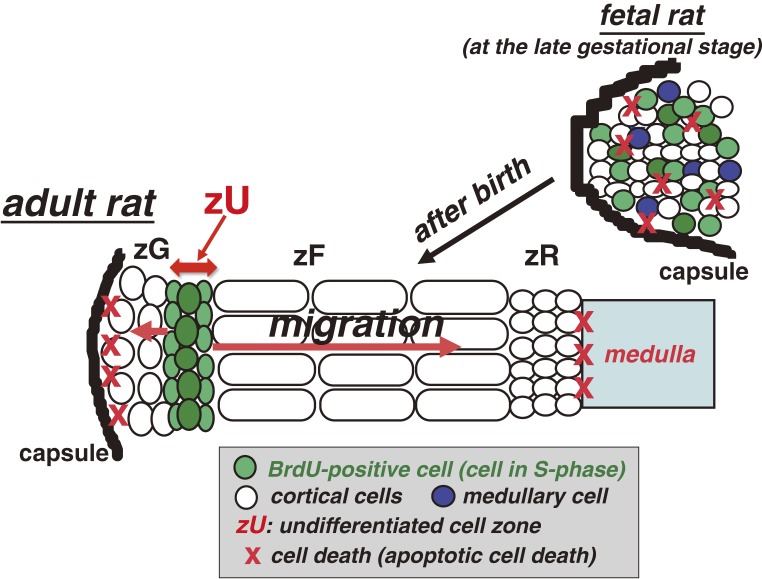
The adrenal cortex of mammals consists of three concentric zones, i.e., the zona glomerulosa (zG), the zona fasciculata (zF), and the zona reticularis (zR), which secrete mineralocorticoids, glucocorticoids, and adrenal androgens, respectively. In 1994, we identified immunohistochemically a new zone between zG and zF of the rat adrenal gland. The zone appeared to be devoid of any significant endocrine functions specific to adrenocortical zones, therefore, we designated the zone as "undifferentiated cell zone (zU)". Further, BrdU (5-bromo-2'-deoxyuridine)-incorporating cells (cells in S-phase) were concentrated at the outer region and the inner region of zU, and these cells proliferated and migrated bidirectionally: toward zG centrifugally and toward zF centripetally. We proposed that cells in and around zU are stem/progenitor cells of the rat adrenal cortex, maintaining functional zonation of the adrenal cortex. The view is consistent with observations reported recently that Sonic hedgehog (Shh), an important factor in embryonic development and adult stem cell maintenance, exists in zU of the rat adrenal gland and the Shh-containing cells seem to migrate bidirectionally.


Figures

Figure 1.
Figure 1.
Effect of concentration of cholate on the solubilization of P450 from sonicated bovine adrenocortical mitochondria. After incubation of sonicated mitochondria with Na-cholate, the samples were centrifuged at 105,000 × ɡ for 90 min and the supernatants were analyzed. —○—: P450; Δ Absorbance between 450 and 490 nm in the carbon monoxide difference spectrum. – –●– –: P420; Δ Absorbance between peak (420–423 nm) and trough (430–433 nm) in the carbon monoxide difference spectrum. · · · · × · · · ·: Concentration of protein. Data from Mitani and Horie.43)
Figure 2.
Figure 2.
Effect of steroid on the absolute absorption spectrum of the oxidized form of partially purified bovine mitochondrial P450 preparation. ——: control without addition of steroid. · · · · ·: with deoxycorticosterone (about 32 µM). – – –: with pregnenolone (about 32 µM). Data from Mitani and Horie.43)
Figure 3.
Figure 3.
Effect of adrenodoxin concentration on deoxycorticosterone 11β-hydroxylation in the reconstituted system. (A) 11β-hydroxylase reactions at various concentrations of adrenodoxin were carried out in the reconstituted 11β-hydroxylase system with 1 µM (● — ●) or 2 µM (○ — ○) of partially purified cytochrome P450. (B) The reactions were performed at a fixed concentration (94 µM) of adrenodoxin (○ — ○) or at a fixed molar ratio of adrenodoxin to P450 (adrenodoxin to P450 = 127) (● - - - - ●) with various concentrations of P450. AdX: adrenodoxin. Data from Mitani et al.49)
Figure 4.
Figure 4.
Biosynthetic pathway of steroid hormones in the adrenal cortex taking cholesterol as the starting material. The gray area shows the pathway of aldosterone formation from deoxycorticosterone catalyzed by CYP11B2. Corticosterone and 18-hydroxycorticosterone are the intermediates. Reactions in the square proceed in humans and some other mammals, but not in the adrenal cortex of most rodents due to the lack of CYP17 in the adrenal cortex. zG: zona glomerulosa, zFR: zonae fasciculata-reticularis, zR: zona reticularis.
Figure 5.
Figure 5.
SDS-PAGE analysis of CYP11B2 and CYP11B1 purified from rat adrenal gland. The bands were visualized by silver staining. Apparent molecular weights of CYP11B2 and CYP11B1 determined from the comparison of the protein markers were 49.5 kDa and 51.5 kDa, respectively. Data from Ogishima et al.75)
Figure 6.
Figure 6.
Electron microscopic view of the immunocytochemical staining for CYP11B1 localization in a zF cell of bovine adrenal gland. The section was treated directly with peroxidase-labeled anti-bovine CYP11B1 (Fab′). CYP11B1 (in black) is located mainly on the surfaces of the inner mitochondrial membranes along with tubulovesicular cristae. Data from Mitani et al.77)
Figure 7.
Figure 7.
Localization of CYP11B2 and CYP11B1 in the rat adrenal gland by immunohistochemical staining. A: An adrenal section from a rat fed on a control diet stained with the anti-rat CYP11B2 and the anti-rat CYP11B1 antibodies simultaneously. B: An adrenal section from a rat fed on a Na-deficient diet for 20 days stained with the anti-rat CYP11B2 and the anti-rat CYP11B1 antibodies simultaneously. In both figures, blue and brown colors show the presence of CYP11B2 and CYP11B1, respectively. Nuclei were poststained with methyl green. zG: zona glomerulosa, zF: zona fasciculata, zR: zona reticularis. Data from Mitani et al.81)
Figure 8.
Figure 8.
Histological features of the undifferentiated cell zone (zU). The adrenal sections from rats fed on Na-deficient diet for 20 days were used for the staining. A: An adrenal section stained with anti-rat CYP11B2 (in blue) and anti-rat CYP11B1 (in brown) antibodies simultaneously. Nuclei were poststained with methyl green. B: Nuclei in adrenocortical cells stained with hematoxylin (in black). C: Alkaline phosphatase activities in the adrenal cortex visualized enzyme-histochemically to show the localization of microvasculatures (in dark red). zG: zona glomerulosa, zF: zona fasciculata, zU: undifferentiated cell zone. Data from Mitani et al.81)
Figure 9.
Figure 9.
Spatial distribution of BrdU-positive cells and the movement in the adrenal cortex. (A) Spatial distribution of BrdU-positive cells in the adrenal cortex. □, BrdU-positive cells in the adrenal cortex from rats fed on a control diet for 10 days and sacrificed at 4:00 a.m. ■, BrdU-positive cells in the adrenal cortex from rats fed on a Na-deficient diet for 10 days and sacrificed at 8:00 p.m. In both rats, BrdU was injected at 1 h before sacrifice. (B) Behavior of BrdU-positive cells under Na-deficiency. Rats fed on a Na-deficient diet for 10 days were injected BrdU at 7:00 p.m. and sacrificed at 1 h (gray column) or at the 20th day (black column) after the injection. *P < 0.05, **P < 0.01, ***P < 0.001: data obtained at 1 h vs. at the 20th day in a comparable region. Numbers on the X-axis (relative distance from 0 edge) show the distance (cell layer) from “0”, which is defined as the position of the innermost cell layer of the undifferentiated cell zone (zU). Values are mean ± SEM. zG: zona glomerulosa, zF: zona fasciculata, zU: undifferentiated cell zone. Data from Miyamoto et al.88,89) and Mitani et al.90)
Figure 10.
Figure 10.
Circadian rhythm in the cell proliferation in the adrenal cortex during a 24-hours period. (A) Circadian rhythm in the cell proliferation in zF of rats fed on either control (□) or Na-deficient (■) diets for 10 days. The total numbers of BrdU-positive cells/section were plotted as a function of the time of a day. ACTH concentrations (● - - - - ●) in plasma of rats fed on a control diet were also plotted as a function of the time of a day. (B) Circadian rhythm in the cell proliferation in zG of rats fed on either control (□) or Na-deficient (■) diets for 10 days. The total numbers of BrdU-positive cells/section were plotted as a function of the time of a day. Values are mean ± SEM. (**p < 0.01; data on Na-deficient rats vs control rat at 8:00 p.m.). Refer to data from Miyamoto et al.,88,89) and Mitani et al.90)
Figure 11.
Figure 11.
Capsular and decapsular portion after the enucleation of the adrenal gland. The enucleation of the adrenal gland was performed using rats fed on Na-deficient diet for 20 days in order to detect zG clearly as CYP11B2-expressing zone. After the enucleation, the capsular and the decapsular portion were stained using anti-rat CYP11B2 and anti-rat CYP11B1 antibodies simultaneously. A: The capsular portion of the adrenal gland. Note that, on the plate, a section of the capsular portion is bent, and one inner edge of zU is located side by side with another edge of zU. B: The decapsular portion of the adrenal gland. In both figures, blue and brown colors show the presence of CYP11B2 and CYP11B1, respectively. Nuclei were poststained with methyl green. zG: zona glomerulosa, zF: zona fasciculata, zR: zona reticularis, zU: undifferentiated cell zone. Refer to data from Mitani et al.100) and Mitani and Ishimura.101)
Figure 12.
Figure 12.
Proposed mechanism on the development and maintenance of the functional zonation of the rat adrenal cortex. In fetal adrenal gland, medullary and cortical cells (mostly zF cells) are intermingled. BrdU-positive cells (cells in S-phase) are scattered over the gland and proliferate without apparent movement. After birth, the cortical zonation becomes distinct with zU. At the mature stage, BrdU-positive cells are concentrated in and around the zU and migrate bidirectionally. Cells differentiating to zF cells migrate centripetally and then transform into zR cells. They finally degenerate at the boundary between zR and the medulla. Meanwhile cells differentiating into zG cells migrate centrifugally and may degenerate within zG. zG: zona glomerulosa, zF: zona fasciculata, zR: zona reticularis, zU: undifferentiated cell zone.

References

    1. Eustachi, B. (1563) Opuscula Anatomica, Vincentius Luchinus, Venice.
    1. Addison, T. (1855) On the constitutional and local effect of disease of the supra-renal capsules. Samuel Highley, London.
    1. Dale H. (1949) Thomas Addison: Pioneer of endocrinology. British Med. J., Aug. 13, 347–352 - PMC - PubMed
    1. Orth, D.N., Kavacs, W.J. and Rowan, D.C. (1992) The adreal cortex. In Wilson’s Textbook of Endocrinology (eds. Wilson, J.D. and Foster, D.W.). Sanders, Philadelphia, pp. 489–619.
    1. Arnold J. (1866) Ein Beitrag zür feineren Struktur und dem Chemismus der Nebennieren. Arch. Pathol. Anat. Physiol. Klin. Med. 35, 64–107

Publication types