. 2014 May 12:4:4920.
doi: 10.1038/srep04920.

Hematopoietic not systemic impairment of Roquin expression accounts for intestinal inflammation in Roquin-deficient mice

Affiliations

Hematopoietic not systemic impairment of Roquin expression accounts for intestinal inflammation in Roquin-deficient mice

Dina Montufar-Solis et al. Sci Rep. .

Abstract

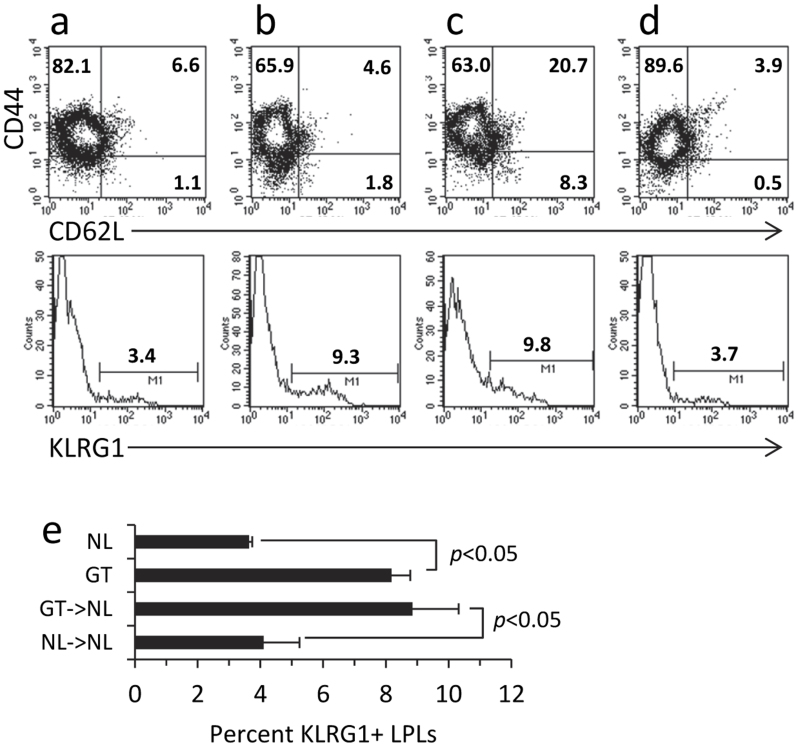
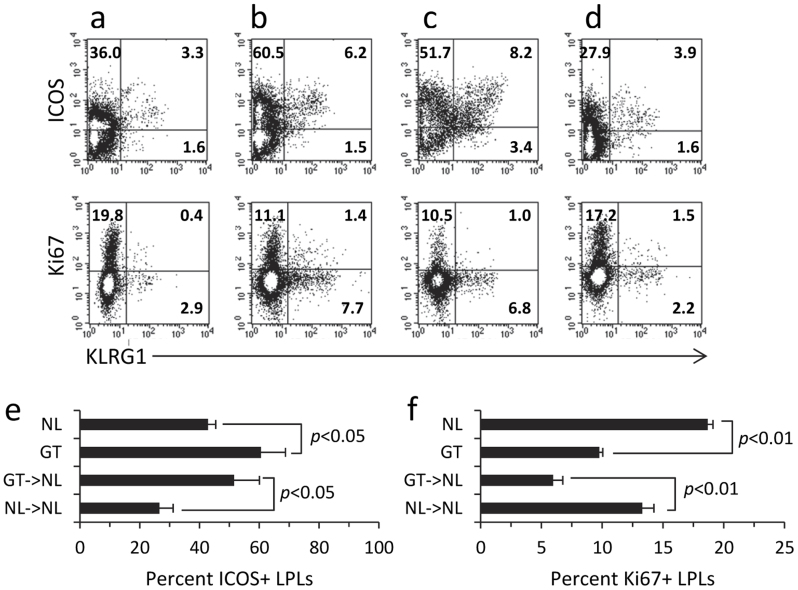
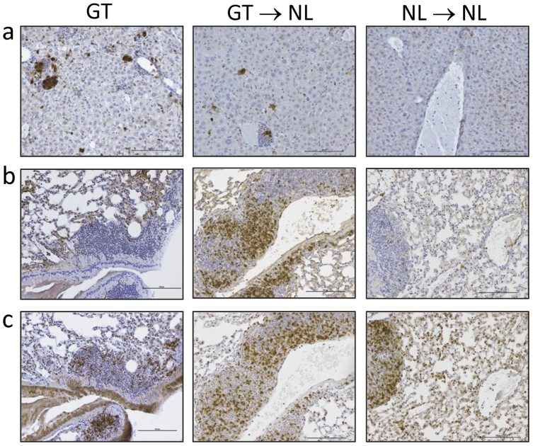
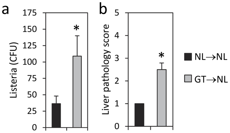
Roquin, an E3 ligase, is involved in curtailing autoimmune pathology as seen from studies using mice with mutated (Rc3h1(san/san)) or disrupted (Rc3h1(gt/gt)) Rc3h1 gene. The extent to which intestinal immunopathology is caused by insufficient Roquin expression in the immune system, or by Roquin impairment in non-hematopoietic cells, has not been determined. Using bone marrow cells from Rc3h1(gt/gt) mice transferred into irradiated normal mice (Rc3h1(gt/gt) → NL chimeras), we show that inflammation developed in the small intestine, kidney, lung, liver, and spleen. Proinflammatory cytokine levels were elevated in lamina propria lymphocytes (LPLs). Inflammation in the liver was accompanied by areas of hepatocyte apoptosis. Lung inflammation consisted of an influx of both T cells and B cells. Small intestinal LPLs had increased numbers of CD44(hi), CD62L(lo), KLRG1(+), ICOS(+) short-lived effector cells, indicating an influx of activated T cells. Following oral infection with L. monocytogenes, Rc3h1(gt/gt) → NL chimeras had more liver pathology and greater numbers of bacteria in the Peyer's patches than NL → NL chimeras. These findings demonstrate that small intestinal inflammation in Rc3h1(san/san) and Rc3h1(gt/gt) mice is due to a failure of Roquin expression in the immune system and not to insufficient systemic Roquin expression.


Figures

Figure 1
Figure 1
Histopathological analysis of intestinal tissue sections from (a) Rc3h1gt/gt mice, (b) Rc3h1gt/gt → NL chimeras, and (c) NL → NL chimeras for the duodenum, jejunum, ileum, and cecum. Note the areas of inflammation (boxed regions) in intestinal tissues of Rc3h1gt/gt and Rc3h1gt/gt → NL chimeras. Micrographs are 100 × original magnification; scale bars are 100 μm. (d) Mean pathology scores ± SEM of 8 Rc3h1gt/gt mice, 5 Rc3h1gt/gt → NL chimeras, and 4 NL → NL chimeras. *p < 0.05, ♦p < 0.01 compared to NL → NL chimeras. (e) Mean numbers of small intestine LPLs ± SEM of 5 Rc3h1gt/gt → NL chimeras and 4 NL → NL chimeras. *p < 0.05 compared to NL → NL chimeras.
Figure 2
Figure 2. The proinflammatory cytokines, IL-17A, IFNγ, and TNFα, are produced at higher levels in LPLs of Rc3h1gt/gt → NL chimeras compared to NL → NL chimeras.
Two-color intracellular staining of LPLs from (a) NL → NL chimeras and (b) Rc3h1gt/gt → NL chimeras. (c) Percent of LPLs producing IL-17A, IFNγ, and TNFα in NL → NL chimeras vs. Rc3h1gt/gt → NL chimeras. Association of cytokine-secreting cells with (d) CD8 LPLs and (e) CD8+ LPLs. *p < 0.05. Mean values ± SEM of 3–5 mice per group.
Figure 3
Figure 3
CD44, CD62L, KLRG1 expression on LPLs from (a) NL mice, (b) Rc3h1gt/gt mice (c) Rc3h1gt/gt → NL chimeras, and (d) NL → NL chimeras. Determination of KLRG1 expression was done by gating onto the CD44hi CD62Llo population. (e) There were proportionally greater numbers of LPLs from Rc3h1gt/gt mice and Rc3h1gt/gt → NL chimeras expressing KLRG1 compared to their respective controls (p < 0.05). Mean values ± SEM of 2–3 samples per group.
Figure 4
Figure 4
KLRG1+ cells in (a) NL mice, (b) Rc3h1gt/gt mice (c) Rc3h1gt/gt → NL chimeras, and (d) NL → NL chimeras are predominantly ICOS+ (top panel), and are Ki67 cells, indicating that they are activated non-proliferating cells. (e) ICOS is expressed on proportionally more LPLs from Rc3h1gt/gt mice and Rc3h1gt/gt → NL chimeras compared to their respective controls (p < 0.05); mean values ± SEM of 2–3 samples per group. (f) LPLs from Rc3h1gt/gt mice and Rc3h1gt/gt → NL chimeras have proportionally fewer Ki67+ proliferating cells compared to their respective controls (p < 0.01). Mean values ± SEM of 2–3 samples per group.
Figure 5
Figure 5
Histopathological analysis of tissue sections from the kidney, lung, liver, and spleen from (a) Rc3h1gt/gt mice, (b) Rc3h1gt/gt → NL chimeras, and (c) NL → NL chimeras. Inflammation was most common in the liver of Rc3h1gt/gt and Rc3h1gt/gt → NL chimeras, and was less common in the kidney, lung, and spleen. Inflammation was absent in the kidney, lung, and liver of NL → NL chimeras. Micrographs are 100 × original magnification; scale bars are 100 μm. (d) Mean liver scores ± SEM of 10 Rc3h1gt/gt mice and 8 Rc3h1gt/gt → NL chimeras. *p < 0.05 comparing Rc3h1gt/gt mice and Rc3h1gt/gt → NL chimeras. (e) Percent of mice with inflammation in the liver, kidney, spleen, lung, and ileum of Rc3h1gt/gt mice compared to Rc3h1gt/gt → NL chimeras.
Figure 6
Figure 6
(a) Caspase 3 expression in the liver of Rc3h1gt/gt mice (GT), Rc3h1gt/gt → NL chimeras (GT → NL), and NL → NL chimeras, showing increased caspase staining in mice with ablated Rc3h1. Lung tissues in the three groups of mice stained for the presence of (b) CD20+ B cells and (c) CD3+ T cells. Note the abundance of B cells in mice with ablated Rc3h1 compared to lung tissue from control mice. Micrographs are 100 × original magnification; scale bars are 100 μm.
Figure 7
Figure 7. Effect of oral infection in Rc3h1gt/gt → NL chimeras.
Rc3h1gt/gt → NL chimeras and NL → NL chimeras were infected orally with L. monocytogenes as described in the Methods. Rc3h1gt/gt → NL chimeras had (a) more bacteria in the Peyer's patches, and (b) more liver pathology than NL → NL chimeras. *p < 0.05. Mean values ± SEM of 3 mice per group.

References

    1. Ciechanover A. The ubiquitin-proteasome pathway: on protein death and cell life. Embo J 17, 7151–7160 (1998). - PMC - PubMed
    1. Lin A. E. & Mak T. W. The role of E3 ligases in autoimmunity and the regulation of autoreactive T cells. Curr Opin Immunol 19, 665–673 (2007). - PubMed
    1. Vinuesa C. G. et al. A RING-type ubiquitin ligase family member required to repress follicular helper T cells and autoimmunity. Nature 435, 452–458 (2005). - PubMed
    1. Yu D. et al. Roquin represses autoimmunity by limiting inducible T-cell co-stimulator messenger RNA. Nature 450, 299–303 (2007). - PubMed
    1. Heissmeyer V. & Vogel K. U. Molecular control of Tfh-cell differentiation by Roquin family proteins. Immunol Rev 253, 273–289 (2013). - PubMed

Publication types

MeSH terms

LinkOut - more resources