

Clinicopathological analysis of 17 primary cutaneous T-cell lymphoma of the γδ phenotype from Japan
- PMID: 24815502
- PMCID: PMC4317912
- DOI: 10.1111/cas.12439
Clinicopathological analysis of 17 primary cutaneous T-cell lymphoma of the γδ phenotype from Japan
Abstract
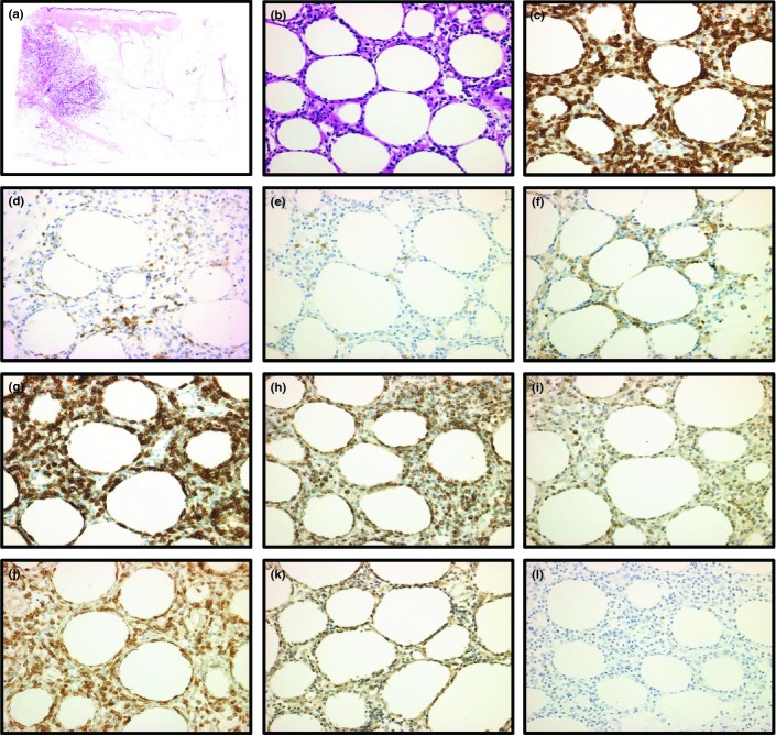
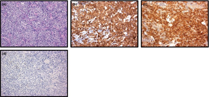
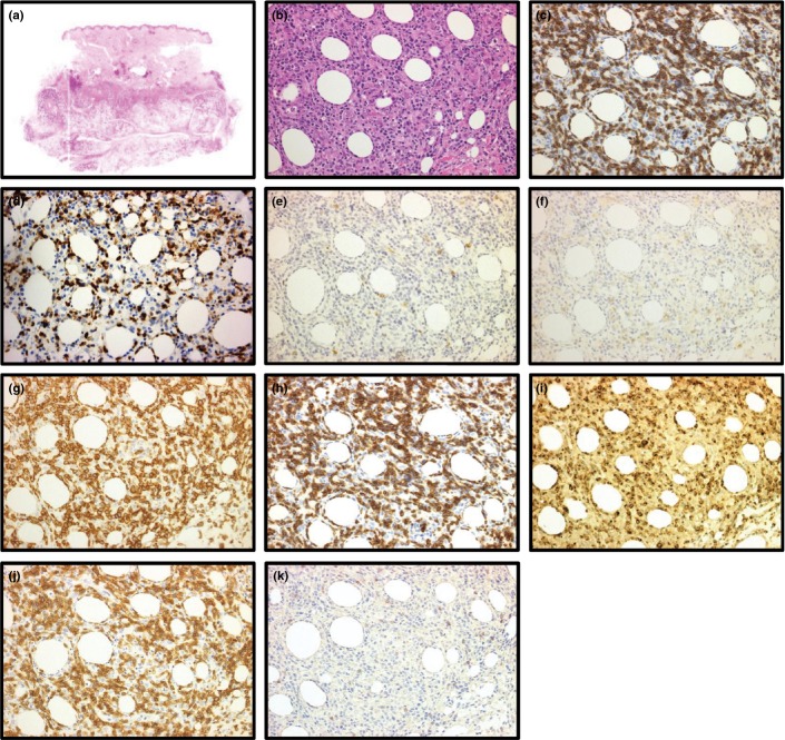
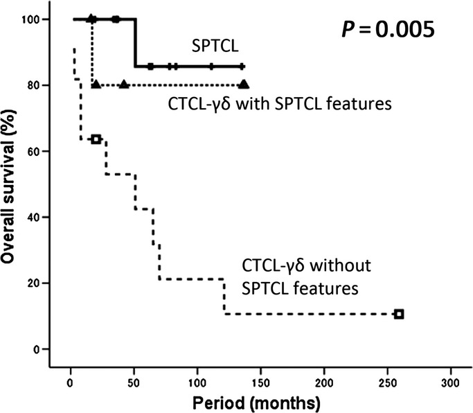
Primary cutaneous γδ T-cell lymphoma (PCGD-TCL) is an aggressive lymphoma consisting of clonal proliferation of mature activated γδ T-cells of a cytotoxic phenotype. Because primary cutaneous γδ T-cell lymphoma is a rare disease, there are few clinicopathological studies. In addition, T-cell receptor (TCR) γδ cells are typically immunostained in frozen sections or determined by TCRβ negativity. We retrospectively analyzed 17 primary cutaneous T-cell lymphomas of the γδ phenotype (CTCL-γδ) in a clinicopathological and molecular study using paraffin-embedded sections. Among 17 patients, 11 had CTCL-γδ without subcutaneous panniculitis-like T-cell lymphoma (SPTCL) features and six had CTCL-γδ with SPTCL features. Immunophenotypically, some significant differences were found in CD8 and CD56 positivity between our patient series of CTCL-γδ patients with SPTCL features and SPTCL-γδ patients described in the previous literature. A univariate analysis of 17 CTCL-γδ patients showed that being more than 60 years old, presence of visceral organ involvement, and small-to-medium cell size were poor prognostic factors. In addition, the 5-year overall survival rate was 42.4% for the CTCL-γδ patients without SPTCL features and 80.0% for those with SPTCL features. Consequently, there was a strikingly significant difference in overall survival among SPTCL, CTCL-γδ with SPTCL features and CTCL-γδ without SPTCL features (P = 0.0005). Our data suggests that an indolent subgroup may exist in CTCL-γδ. Studies on more cases, including those from other countries, are warranted to delineate the clinicopathological features and the significance in these rare lymphomas.
Keywords: Cutaneous γδ T-cell lymphoma; T-cell receptor αβ; T-cell receptor γδ; indolent clinical behavior; subcutaneous panniculitis-like T-cell features.
© 2014 The Authors. Cancer Science published by Wiley Publishing Asia Pty Ltd on behalf of Japanese Cancer Association.
Figures
References
-
- Swerdlow SH, Campo E, Harris HL, et al. WHO Classification of Tumours of Haematopoietic and Lymphoid Tissues. Lyon: IARC; 2008.
-
- Toro JR, Liewehr DJ, Pabby N, et al. Gamma-delta T-cell phenotype is associated with significantly decreased survival in cutaneous T-cell lymphoma. Blood. 2003;101:3407–12. - PubMed
-
- Willemze R, Jaffe ES, Burg G, et al. WHO-EORTC classification for cutaneous lymphomas. Blood. 2005;105:3768–85. - PubMed
-
- Macon WR, Levy NB, Kurtin PJ, et al. Hepatosplenic alpha beta T-cell lymphomas – a report of 14 cases and comparison with hepatosplenic gamma delta T-cell lymphomas. Am J Surg Pathol. 2001;25:285–96. - PubMed
-
- Chan JKC, Chan ACL, Cheuk W, et al. Type II enteropathy-associated T-cell lymphoma: a distinct aggressive lymphoma with frequent gamma delta T-cell receptor expression. Am J Surg Pathol. 2011;35:1557–69. - PubMed
Publication types
MeSH terms
Substances
LinkOut - more resources
Full Text Sources
Other Literature Sources
Medical
Research Materials
