

Endoscopic findings using narrow-band imaging to distinguish between basal cell hyperplasia and carcinoma of the pharynx
- PMID: 24815674
- PMCID: PMC4317933
- DOI: 10.1111/cas.12440
Endoscopic findings using narrow-band imaging to distinguish between basal cell hyperplasia and carcinoma of the pharynx
Abstract
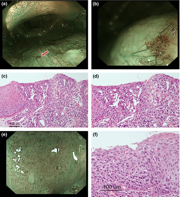
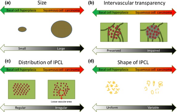
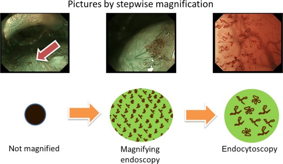
Narrow-band imaging (NBI) has been reported to be useful for detecting superficial-type esophageal or head and neck squamous cell carcinoma (SCC), and in the present study we have used NBI to detect non-carcinomatous lesions, such as basal cell hyperplasia (BCH) accompanied by microvascular irregularities; these non-carcinomatous lesions were pathologically discriminated from squamous cell carcinoma of the pharynx. The aim of the present study was to clarify the endoscopic characteristics of BCH that contribute to the discrimination of superficial-type head and neck SCC (HNSCC). We examined the key endoscopic findings capable of distinguishing BCH from SCC using 26 BCH and 37 superficial-type SCC of the pharynx that had been pathologically diagnosed at our institution between January 2008 and July 2012. The clinicopathological factors were also compared. The size of the BCH lesions was significantly smaller (P < 0.001), and their intervascular transparency was more clearly observed (P < 0.001). Intra-epithelial papillary capillary loop (IPCL) shapes were less variable and monotonous (P < 0.001), and the distribution of the IPCL was more regular with an interval comparable to that of SCC (P < 0.001), although no significant differences in the sharpness of the lesion border, dilatation of IPCL and tortuosity of the IPCL were seen between the BCH and SCC lesions. This study revealed that BCH was an independent entity in terms of not only pathological findings, but also endoscopic findings observed using NBI, such as the regular distribution of IPCL and the preserved intervascular transparency.
Keywords: Basal cell hyperplasia; head and neck cancer; intra-epithelial papillary capillary loop; narrow-band imaging; pharynx.
© 2014 The Authors. Cancer Science published by Wiley Publishing Asia Pty Ltd on behalf of Japanese Cancer Association.
Figures
References
-
- Yoshida T, Inoue H, Usui S, Satodate H, Fukami N, Kudo SE. Narrow-band imaging system with magnifying endoscopy for superficial esophageal lesions. Gastrointest Endosc. 2004;59:288–95. - PubMed
-
- Ishihara R, Inoue T, Uedo N, et al. Significance of each narrow-band imaging finding in diagnosing squamous mucosal high-grade neoplasia of the esophagus. J Gastroenterol Hepatol. 2010;25:1410–5. - PubMed
-
- Takenaka R, Kawahara Y, Okada H, et al. Narrow-band imaging provides reliable screening for esophageal malignancy in patients with head and neck cancers. Am J Gastroenterol. 2009;104:2942–8. - PubMed
-
- Lee CT, Chang CY, Lee YC, et al. Narrow-band imaging with magnifying endoscopy for the screening of esophageal cancer in patients with primary head and neck cancers. Endoscopy. 2010;42:613–9. - PubMed
Publication types
MeSH terms
LinkOut - more resources
Full Text Sources
Other Literature Sources
Medical
Research Materials
