. 2014 May 12:4:4924.
doi: 10.1038/srep04924.

Polarization-sensitive hyperspectral imaging in vivo: a multimode dermoscope for skin analysis

Affiliations

Polarization-sensitive hyperspectral imaging in vivo: a multimode dermoscope for skin analysis

Fartash Vasefi et al. Sci Rep. .

Abstract

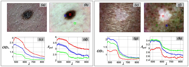
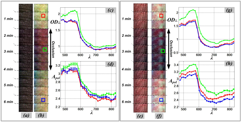
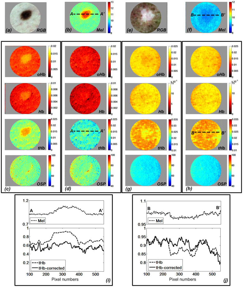
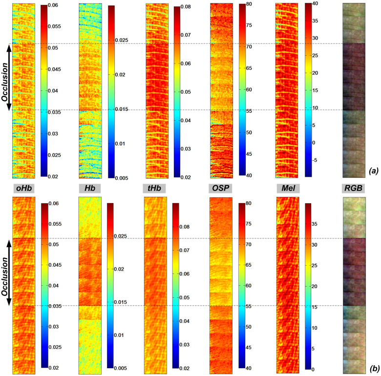
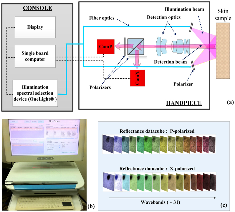
Attempts to understand the changes in the structure and physiology of human skin abnormalities by non-invasive optical imaging are aided by spectroscopic methods that quantify, at the molecular level, variations in tissue oxygenation and melanin distribution. However, current commercial and research systems to map hemoglobin and melanin do not correlate well with pathology for pigmented lesions or darker skin. We developed a multimode dermoscope that combines polarization and hyperspectral imaging with an efficient analytical model to map the distribution of specific skin bio-molecules. This corrects for the melanin-hemoglobin misestimation common to other systems, without resorting to complex and computationally intensive tissue optical models. For this system's proof of concept, human skin measurements on melanocytic nevus, vitiligo, and venous occlusion conditions were performed in volunteers. The resulting molecular distribution maps matched physiological and anatomical expectations, confirming a technologic approach that can be applied to next generation dermoscopes and having biological plausibility that is likely to appeal to dermatologists.


Conflict of interest statement

R.B.S. and A.J.D. declare no competing interests. F.V. is a Research Scientist for SMI. N.M. and R.C. are Consultants for SMI. D.L.F. is CEO and Chairman of SMI. E.H.L. was the President of SMI in the past. SMI is a for-profit corporate entity that intends to use the multimode dermoscope and approach described in the manuscript for early diagnosis of melanoma, and commercialize the device, after securing regulatory approval for it.

Figures

Figure 1
Figure 1. SkinSpect skin images and spectra of melanocytic nevus and vitiligo.
(a) parallel-polarized color image of skin with nevus; (b) cross-polarized color image of skin with nevus; (c) Cross polarized optical density spectra, OD, of nevus core (red), nevus halo (blue), and normal skin (green), (d) Polarized attenuation spectra, Apol, of nevus core, nevus halo, and normal skin; (e) parallel-polarized color image of skin with vitiligo; (f) cross-polarized color image of skin with vitiligo (g) Cross polarized optical density spectra, OD, of vitiligo (red) and normal skin (blue and green), (h) Polarized attenuation spectra, Apol, of vitiligo and normal skin.
Figure 2
Figure 2. SkinSpect skin images (time sequence) and spectra of same anatomical region of interest during venous occlusion.
(a) Color parallel-polarized image sequence and (b) Color cross-polarized image sequence of dorsal-side finger; (c) Cross polarized optical density spectra, OD, and (d) Polarized Attenuation spectra, Apol, of dorsal-side finger; (e) Color parallel-polarized image and (f) Color cross-polarized image of volar-side finger; (g) Cross polarized optical density spectra, OD, and (h) Polarized Attenuation spectra, Apol, of volar-side finger. (Red line: pre-occlusion, green line: during occlusion, blue line: post-occlusion).
Figure 3
Figure 3. Calculated relative skin chromophore distribution of melanocytic nevus and vitiligo showing reduction of melanin-hemoglobin misestimation.
(a) Color cross-polarized image of nevus; (b) relative melanin (Mel); oxy-hemoglobin (oHb), deoxy-hemoglobin (Hb), total hemoglobin (tHb), and Oxygen Saturation Percentage (OSP) distribution for skin with nevus (c) before melanin correction, (d) after melanin correction; (e) Color cross-polarized image of vitiligo; (f) relative melanin (Mel), oxy-hemoglobin (oHb), deoxy-hemoglobin (Hb), total hemoglobin (tHb), and Oxygen Saturation Percentage (OSP) map of skin with vitiligo (g) before melanin correction, (h) after melanin correction (i) relative melanin, total hemoglobin concentration profile at A-A′ in melanocytic nevus (j) relative melanin, total hemoglobin concentration profile at B-B′ in vitiligo.
Figure 4
Figure 4. Relative molar absorptivity of Oxy-hemoglobin (oHb), deoxy-hemoglobin (Hb), total hemoglobin (tHb), melanin and oxygen saturation (OSP) m aps with corresponding color cross-polarized image of (a) dorsal finger and (b) volar finger during finger cuff occlusion.
Figure 5
Figure 5
(a) SkinSpect research prototype system diagram and the (b) SkinSpect console; (c) SkinSpect data output includes reflection images over the range from 467 nm to 857 nm, in parallel and cross polarization modes.
Figure 6
Figure 6
(a) Skin mole boundaries determined from the grayscale image at 500 nm (white) compared to the image at 700 nm (green); (b) resolution target boundaries determined from the grayscale image at 500 nm (white) compared to the image at 700 nm (green); (c) skin mole boundaries determined from the successive scans (typical, not best result); (d) image registration between parallel and cross polarization images.

References

    1. Kollias N. & Stamatas G. Optical non-invasive approaches to diagnosis of skin diseases. J Invest Derm Symp P 7, 64–75 (2002). - PubMed
    1. Elbaum M. et al. Automatic differentiation of melanoma from melanocytic nevi with multispectral digital dermoscopy: a feasibility study. J Am Acad Dermatol 44, 207–18 (2001). - PubMed
    1. Monheit G. et al. The performance of MelaFind: a prospective multicenter study. Arch Dermatol 147, 188–94 (2011). - PubMed
    1. Kupetsky E. A. & Ferris L. K. The diagnostic evaluation of MelaFind multi-spectral objective computer vision system. Expert Opinion on Medical Diagnostics 0, 1–7 (2013). - PubMed
    1. Gutkowicz-Krusin D. et al. Precision of automatic measurements of pigmented skin lesion parameters with a MelaFind multispectral digital dermoscope. Melanoma Res 10, 563–70 (2000). - PubMed

Publication types