. 2014 May;70(Pt 5):1321-35.
doi: 10.1107/S1399004714002739. Epub 2014 Apr 30.

Structure of sulfamidase provides insight into the molecular pathology of mucopolysaccharidosis IIIA

Affiliations

Structure of sulfamidase provides insight into the molecular pathology of mucopolysaccharidosis IIIA

Navdeep S Sidhu et al. Acta Crystallogr D Biol Crystallogr. 2014 May.

Abstract

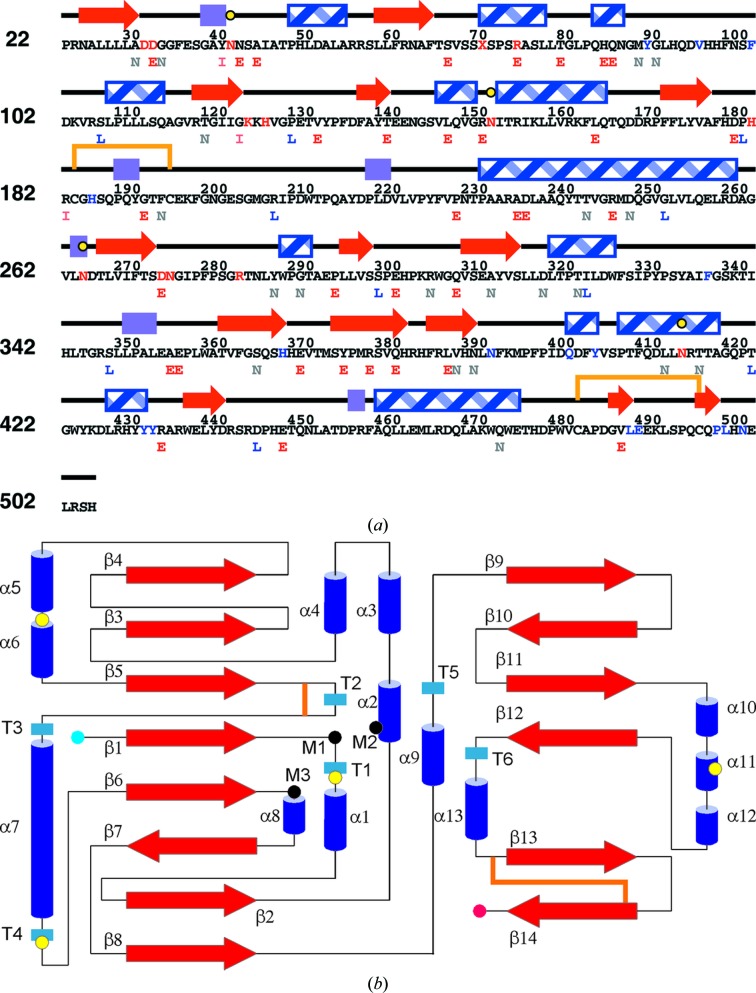
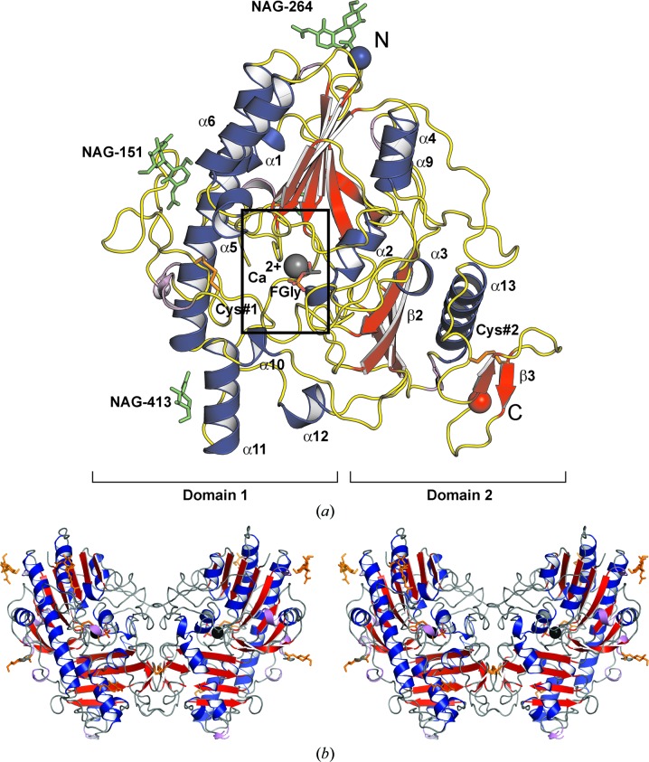
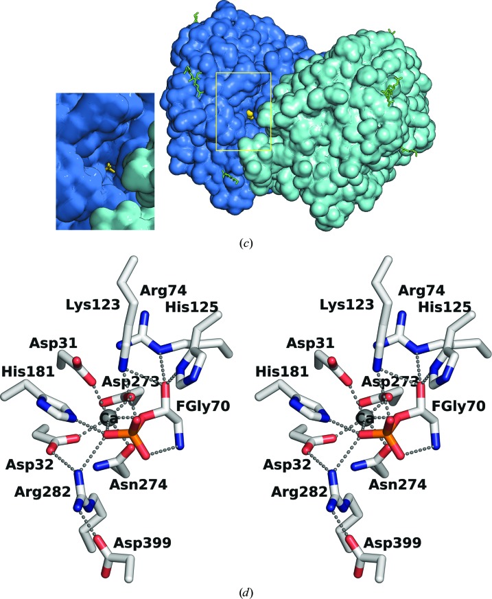
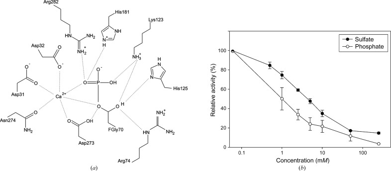
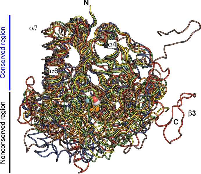
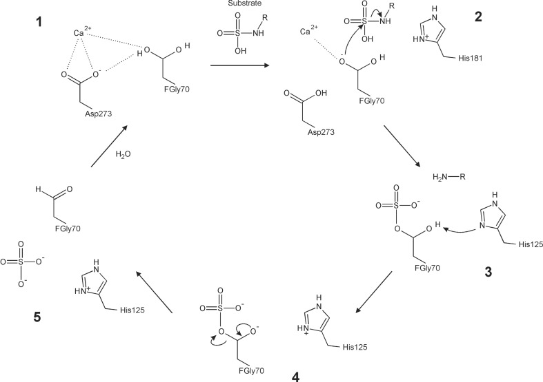
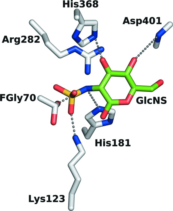
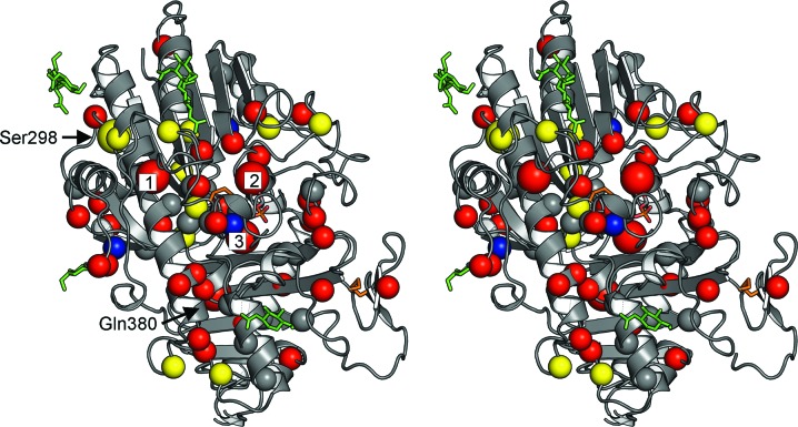
Mucopolysaccharidosis type IIIA (Sanfilippo A syndrome), a fatal childhood-onset neurodegenerative disease with mild facial, visceral and skeletal abnormalities, is caused by an inherited deficiency of the enzyme N-sulfoglucosamine sulfohydrolase (SGSH; sulfamidase). More than 100 mutations in the SGSH gene have been found to reduce or eliminate its enzymatic activity. However, the molecular understanding of the effect of these mutations has been confined by a lack of structural data for this enzyme. Here, the crystal structure of glycosylated SGSH is presented at 2 Å resolution. Despite the low sequence identity between this unique N-sulfatase and the group of O-sulfatases, they share a similar overall fold and active-site architecture, including a catalytic formylglycine, a divalent metal-binding site and a sulfate-binding site. However, a highly conserved lysine in O-sulfatases is replaced in SGSH by an arginine (Arg282) that is positioned to bind the N-linked sulfate substrate. The structure also provides insight into the diverse effects of pathogenic mutations on SGSH function in mucopolysaccharidosis type IIIA and convincing evidence for the molecular consequences of many missense mutations. Further, the molecular characterization of SGSH mutations will lay the groundwork for the development of structure-based drug design for this devastating neurodegenerative disorder.

Keywords: mucopolysaccharidosis IIIA; sulfamidase.


Figures

Figure 1
Figure 1
Schematic representations of the SGSH structure. (a) Mapping of SGSH primary and secondary structures. β-Strands, red arrows; α-helices, blue striped rectangles; 310-helices, violet rectangles; the two disulfide bridges are shown as orange lines and the four glycosylated asparagines as yellow filled circles. Functionally important residues (active site and glycosylation sites) are shown in red; some of the residues at the dimer interface are shown in blue. The phenotype of the missense mutation sites is indicated below the sequence as follows: early-onset disease (E), red; intermediate-onset (I), orange; late-onset (L), blue; phenotype not reported in the literature (N), grey. (b) Topology diagram (not drawn to scale). Colour coding is similar to that in Fig. 1 ▶(a), with α-helices shown as blue cylinders and the N-terminus and C-terminus as blue and red circles, respectively. Divalent metal-binding residues are labelled M1 (Asp31, Asp32), M2 (FGly70) and M3 (Asp273, Asn274). Secondary-structure elements are named as indicated in the main text.
Figure 2
Figure 2
Three-dimensional structure of SGSH. (a) Monomer. The approximate locations of domains 1 and 2 are shown (square brackets), with β-sheets in domain 2 labelled β2 and β3. β-Strands are shown in red, α-helices in blue and loops in yellow. Cystine bridges are shown in orange (Cys#1, 183–194; Cys#2, 481–495). The N-terminus (N) is shown as a blue ball and the C-terminus (C) as an orange ball. The formylglycine (FGly) 70 side chain is shown as a stick model in standard colours. The Ca2+ ion is shown as a grey ball. Glycosylation sites (‘NAG-’ followed by the asparagine residue number) are shown as green sticks. (b) Dimer. The dimer noncrystallographic symmetry axis lies vertically in the plane of the paper, with subunit centroids in the approximate paper plane on either side of it. FGly70, cystine bridges and glycosylations are shown as orange stick models. Other representations are as in Figs. 1 ▶ and 2 ▶(a). (c) A short tunnel from a surface cleft leads to the active site. The inset on the left shows an enlargement of the boxed area. The two dimer subunits are shown in blue and cyan, FGly is shown as yellow spheres or sticks and glycosylations as green sticks. (d) Active site as viewed from its entry (stick models; the major interactions shown are described in the main text).
Figure 2
Figure 2
Three-dimensional structure of SGSH. (a) Monomer. The approximate locations of domains 1 and 2 are shown (square brackets), with β-sheets in domain 2 labelled β2 and β3. β-Strands are shown in red, α-helices in blue and loops in yellow. Cystine bridges are shown in orange (Cys#1, 183–194; Cys#2, 481–495). The N-terminus (N) is shown as a blue ball and the C-terminus (C) as an orange ball. The formylglycine (FGly) 70 side chain is shown as a stick model in standard colours. The Ca2+ ion is shown as a grey ball. Glycosylation sites (‘NAG-’ followed by the asparagine residue number) are shown as green sticks. (b) Dimer. The dimer noncrystallographic symmetry axis lies vertically in the plane of the paper, with subunit centroids in the approximate paper plane on either side of it. FGly70, cystine bridges and glycosylations are shown as orange stick models. Other representations are as in Figs. 1 ▶ and 2 ▶(a). (c) A short tunnel from a surface cleft leads to the active site. The inset on the left shows an enlargement of the boxed area. The two dimer subunits are shown in blue and cyan, FGly is shown as yellow spheres or sticks and glycosylations as green sticks. (d) Active site as viewed from its entry (stick models; the major interactions shown are described in the main text).
Figure 3
Figure 3
Active site and enzyme inhibition. (a) Schematic of the active-site region in SGSH. A Ca2+ ion is coordinated by side-chain P atoms from Asp31, Asp32, Asp273, Asn274 and the phosphorylated FGly70, which is in turn stabilized by interactions with the side chains of residues Arg74, Lys123, His125, His181, Asp273 and Arg282. (b) Inhibition of SGSH acitivity by phosphate and sulfate. The IC50 of phosphate was determined to be 1 mM; the IC50 of sulfate was 5 mM.
Figure 4
Figure 4
Superposition of the SGSH backbone on those of five related sulfatases: SGSH (red), ASA (orange), ASB (yellow), PAS (blue), GALNS (green) and BcPMH (brown). SGSH shares a common fold with O-sulfatases consisting of a large central β-sheet with decorating helices (‘conserved region’, top); the loops form a more variable region (‘nonconserved region’, bottom). For orientation, the SGSH N-terminus and C-terminus are shown (N and C, respectively), as are some secondary-structure elements (as in Fig. 2 ▶ a) and some atoms in the active site in ball representation: Ca2+ (dark grey), phosphate O atoms (orange), FGly Cβ (light grey) and free hydroxyl O atom (red).
Figure 5
Figure 5
Proposed reaction mechanism in SGSH (schematic). The active-site formylglycine (FGly70), which is intrinsically reactive, undergoes hydration to form the resting state of the enzyme with a gem-diol group (step 1). Coordination of one of the hydroxyl groups of the gem-diol to a Ca2+ ion facilitates the development of a negative charge on the O atom as its proton is lost to a base. The negatively charged O atom nucleophilically attacks the sulfur centre of the N-linked sulfate group on the glucosamine substrate (step 2), resulting in a covalently bound enzyme–substrate complex with a pentavalent sulfur transition state. An acid (possibly His181) facilitates the cleavage of the S—N bond by protonating the bridging N atom to form an amine leaving group on the N-desulfated substrate, which diffuses away, leaving an O-sulfated enzyme (step 3). Finally, in a step that underlines the importance of the formylglycine residue, another base (His125) deprotonates the second hydroxyl group, resulting in a negatively charged O atom (step 4) that forms a double bond with the Cβ atom as the C—O bond between it and the bridging O atom of the sulfate group breaks, eliminating the sulfate ion and regenerating the formylglycine residue (step 5).
Figure 6
Figure 6
Hypothetical model showing some proposed interactions between the terminal N-sulfoglucosamine residue (GlcNS; C atoms in green, other atoms in standard colours) of the substrate with the enzyme in the active site (C atoms in light grey). His181 acts as the acid facilitating desulfation of the substrate. Other residues that help to bind and orient the substrate include the side chains of FGly70, Lys123, Arg282 and His368 and the main-chain amide N atom of Asp401.
Figure 7
Figure 7
Stereo figure showing missense mutations mapped onto the structure of the SGSH monomer. Cα atoms of residues associated with an early-onset phenotype are shown in red, those associated with an intermediate-onset phenotype in blue and those associated with a late-onset phenotype in yellow. Missense mutations for which the phenotype was not reported are indicated in grey. Most mutations with known phenotype are early-onset mutations. Late-onset mutations appear to map closer to the periphery of the enzyme. Some of the most common mutations are indicated by a larger ball size. These are Ser298, Arg245 (indicated ‘1’), Arg74 (‘2’), Ser66 (‘3’) and Gln380. The orientation shown is the same as for one of the subunits (on the left) of the dimer in Fig. 2 ▶(b); the active site is indicated by FGly70 (stick model; standard colours) and Ca2+ ion (black ball). Glycosylations are shown as green sticks.

Similar articles

Cited by

References

    1. Baehner, F., Schmiedeskamp, C., Krummenauer, F., Miebach, E., Bajbouj, M., Whybra, C., Kohlschütter, A., Kampmann, C. & Beck, M. (2005). J. Inherit. Metab. Dis. 28, 1011–1017. - PubMed
    1. Ballabio, A. & Gieselmann, V. (2009). Biochim. Biophys. Acta, 1793, 684–696. - PubMed
    1. Beesley, C. E., Young, E. P., Vellodi, A. & Winchester, B. G. (2000). J. Med. Genet. 37, 704–707. - PMC - PubMed
    1. Bekri, S., Armana, G., De Ricaud, D., Osenda, M., Maire, I., Van Obberghen, E. & Froissart, R. (2005). J. Inherit. Metab. Dis. 28, 601–602. - PubMed
    1. Bielicki, J., Fuller, M., Guo, X.-H., Morris, C. P., Hopewood, J. J. & Anson, D. S. (1995). Biochem. J. 311, 333–339. - PMC - PubMed

Publication types

Associated data