. 2014 May;70(Pt 5):1375-90.
doi: 10.1107/S1399004714003228. Epub 2014 Apr 30.

An arginine tetrad as mediator of input-dependent and input-independent ATPases in the clock protein KaiC

Affiliations

An arginine tetrad as mediator of input-dependent and input-independent ATPases in the clock protein KaiC

Rekha Pattanayek et al. Acta Crystallogr D Biol Crystallogr. 2014 May.

Abstract

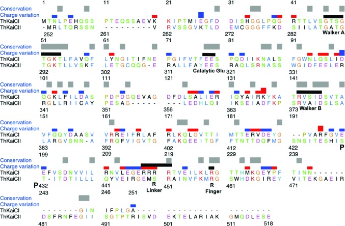
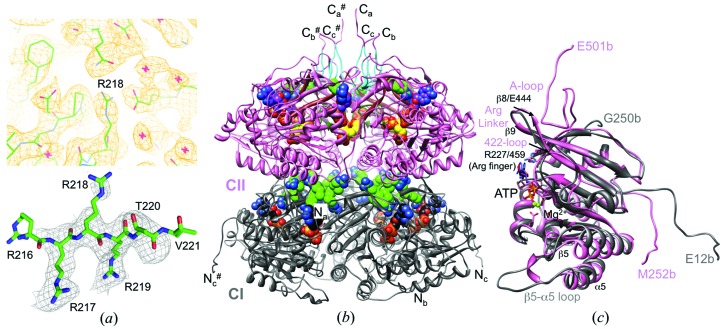
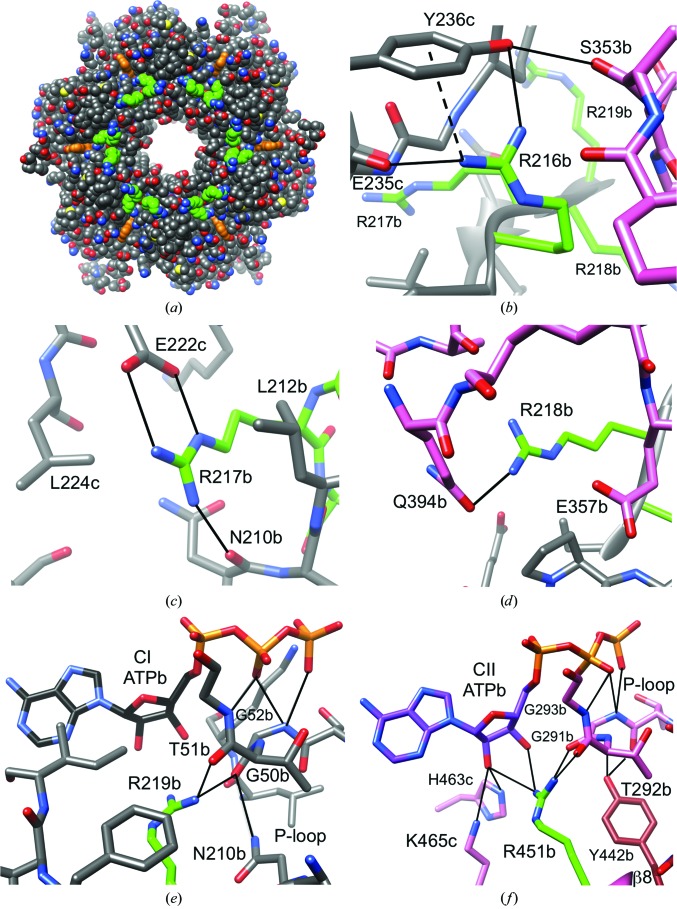
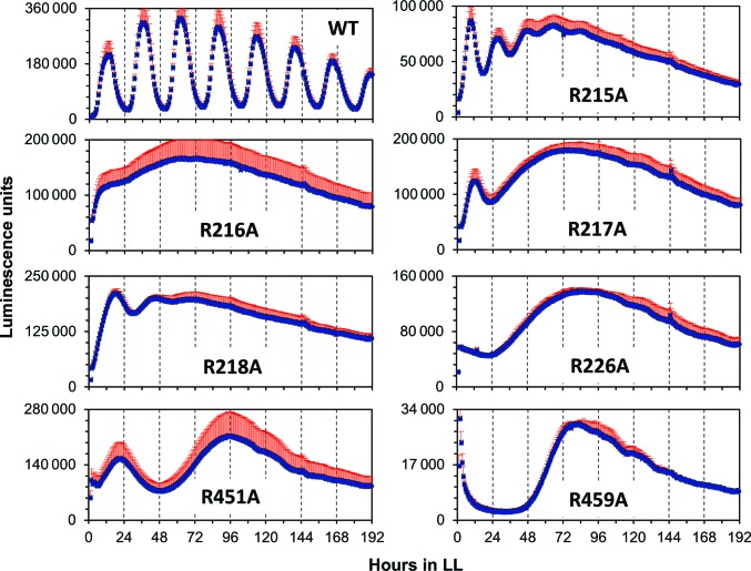
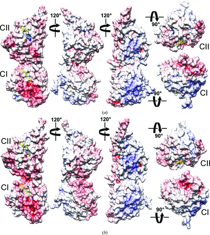
A post-translational oscillator (PTO) composed of the proteins KaiA, KaiB and KaiC is at the heart of the cyanobacterial circadian clock. KaiC interacts with KaiA and KaiB over the daily cycle, and CII domains undergo rhythmic phosphorylation/dephosphorylation with a 24 h period. Both the N-terminal (CI) and C-terminal (CII) rings of KaiC exhibit ATPase activity. The CI ATPase proceeds in an input-independent fashion, but the CII ATPase is subject to metabolic input signals. The crystal structure of KaiC from Thermosynechococcus elongatus allows insight into the different anatomies of the CI and CII ATPases. Four consecutive arginines in CI (Arg linker) that connect the P-loop, CI subunits and CI and CII at the ring interface are primary candidates for the coordination of the CI and CII activities. The mutation of linker residues alters the period or triggers arhythmic behavior. Comparison between the CI and CII structures also reveals differences in loop regions that are key to KaiA and KaiB binding and activation of CII ATPase and kinase. Common packing features in KaiC crystals shed light on the KaiB-KaiC interaction.

Keywords: ATPase; KaiABC; circadian clock; kinase.


Figures

Figure 1
Figure 1
Sequence alignment of the CI (ThKaiCI) and CII (ThKaiCII) domains of T. elongatus KaiC. Conserved residues are indicated by gray bars and changes in the charge of corresponding side chains are marked with blue (CI neutral to CII positive or CI negative to CII neutral) or red bars (CI positive to CII neutral or CI neutral to CII negative). Functionally important residues are indicated by black bars and labeled (the phosphorylation sites Ser431 and Thr432 are marked with a P). Arginine linkers are residues 216–219 in CI and residue 451 in CII. The sequence alignment was generated with Clustal Omega (http://www.ebi.ac.uk/Tools/msa/clustalo/; Sievers et al., 2011 ▸) and modified manually in UCSF Chimera (Pettersen et al., 2004 ▸).
Figure 2
Figure 2
KaiC model quality and overall conformation. (a) Top: quality of the final Fourier 2F oF c sum electron density drawn at the 1σ level in the region of Arg linker residue Arg218. Red crosses represent water molecules. Bottom: example of the quality of the OMIT electron density drawn at the 2.5σ level (residues Arg217–Thr220 were deleted). (b) Ribbon diagram of the overall structure of the T. elongatus KaiC hexamer. Residues of the CI (11–251) and CII (252–500) domains are colored gray and pink, respectively, and the N- and C-termini of subunits are labeled (# indicates a symmetry-related strand). ATPs and arginine linkers (light green C atoms; Arg216–Arg219 in CI and R451 in CII) are highlighted in CPK mode. C atoms of the six ATP molecules bound between the CI and CII subunits are colored black and purple, respectively, and C atoms of the phosphorylated residues Thr432 and Ser431 are colored yellow. A-loop residues (Glu487–Ile497) are cyan, 422-loop residues (Thr415–Ile425) are dark green and β8 strand residues (Thr436–Glu444) are brown. (c) Superimposition of the CI and CII domains from subunit b.
Figure 3
Figure 3
Arginine-linker interactions in the CI and CII halves. (a) Axial view of the 24 CI Arg-linker residues (6 × Arg216–Arg219; green C atoms) located in the waist region of the KaiC hexamer. ATP molecules are highlighted in orange. Interactions are shown for (b) CI Arg216, (c) CI Arg217, (d) CI Arg218, (e) CI Arg219 and (f) CII Arg451. The color code for residues is identical to that in Fig. 2 ▸: C atoms of arginine side chains are highlighted in green, residues from CI domains are gray and those from CII domains are pink. Selected residues are labeled, with a, b and c designating the subunit; the dashed line indicates a cation–π interaction.
Figure 4
Figure 4
Phenotypes of S. elongatus KaiC arginine-linker and arginine-finger alanine mutants. Individual charts show regular rhythm (wild type) and the absence thereof for Arg-linker mutants (CI, R215A, R216A, R217A, R218A; CII, R451A) and Arg-finger mutants (CI, R226A; CII, 459A) under an LL regimen over the course of 8 d as assessed by luminescence. Error bars are shown in red.
Figure 5
Figure 5
KaiCII A-loop conformation and interactions. (a) Conformation of the A-loop, amino acids Glu487–Ile497, in subunit B of T. elongatus KaiC. C atoms of loop residues are highlighted in black and C atoms of additional B-subunit residues, E444b and N443b from the β8 strand, and residues from the adjacent A and C subunits that all interact with the loop are colored pink, brown and pale pink, respectively. Selected side chains are labeled (underlined in the case of loop residues), hydrogen bonds are shown as thin solid lines and hydrophobic contacts are shown as blue dashed lines. (b) Superimposition of the A-loops from the T. elongatus (black C atoms) and S. elongatus (cyan C atoms) KaiC hexamers (B subunit). Notice the very similar conformations and identical sequences with the exception of residue 488 (Gly, T. elongatus, green; Arg, S. elongatus, orange). Hydrogen bonds are shown as thin solid lines and an additional interaction between the side chains of Arg488 and Thr495 in some subunits of S. elongatus KaiC is indicated by a dashed line.
Figure 6
Figure 6
Conformations of the extended phosphorylation site loop (422-loop) in KaiCII and the corresponding region in CI (B subunit). (a) Superimposition of the T. elongatus KaiCII loop region comprising residues Thr415–pThr432 (pink) and the KaiCI loop comprising residues Glu184–Glu199 (gray). Labels indicate equivalent residues based on the sequence alignment of the CI and CII halves (Fig. 1 ▸). The CI loop makes a tighter turn as it lacks amino acids corresponding to Ser422 and Asn423 in CII. CI residues Phe195, Gly196, Val197, Glu198 and Glu199 are part of a short α-helix. The r.m.s.d. for 16 Cα pairs amounts to 2.6 Å. (b) The superimposition of the extended 422-loops in T. elongatus (pink) and S. elongatus KaiC (cyan) reveals similar conformations in spite of deviating sequences in the apical region (Ala422-His423 versus Ser422-Asn423, respectively).
Figure 7
Figure 7
Assessing a putative kinase activity of the KaiCI domain. SDS–PAGE assay of the phosphorylation status of full-length S. elongatus KaiC (KaiC), KaiC treated with λ phosphatase (λPP), S. elongatus KaiCI (wt-KaiCI), the S. elongatus KaiCI triple mutant A192T/E197S/E198T (TST-KaiCI) and S. elongatus KaiCII (KaiCII). Residue numberings between CI from S. elongatus and T. elongatus differ by one, i.e. S. elongatus mutations A192T/E197S/E198T correspond to T. elongatus mutations A193T/E198S/E199T. Multiple bands for KaiC and the double band for KaiCII represent the phosphorylated (top) and nonphosphorylated (bottom) states. In contrast, single bands for wt-­KaiCI and TST-KaiCI indicate that neither protein is phosphorylated. That KaiCII expressed as a separate domain still exhibits phosphorylation despite its inability to form stable hexamers has previously been demonstrated (Pattanayek et al., 2008 ▸).
Figure 8
Figure 8
Similarities between KaiC crystal-packing interactions and KaiC–KaiB binding. Packing interactions between the KaiCII ring and a KaiCI subunit from a symmetry-related KaiC hexamer (KaiC#) in the crystal structures of (a) T. elongatus KaiC and (b) S. elongatus KaiC. (c) The KaiCII–KaiB interaction (only a single KaiB is depicted) in the cryo-EM model of the S. elongatus KaiC6B6 complex. (d, e) The KaiCII–KaiCI# crystal-packing interactions shown in (b) and (c), respectively, rotated by 90° around the horizontal axis and viewed from the side. (f) Superimposition of the latch regions in T. elongatus KaiCI (residues 110–124) and CII (residues 350–356) and the adjacent α-­helical and β-stranded portions.
Figure 9
Figure 9
Electrostatic surface potentials (ESPs) of T. elongatus and S. elongatus KaiC monomers. ESP of (a) T. elongatus KaiC and (b) S. elongatus KaiC (PDB entry 3dvl). KaiC monomeric subunits are depicted in five different orientations, first viewed from the side and related by rotations of 120° around the vertical and then from the top (CII side) and the bottom (CI side). The minimum and maximum values of the electrostatic potential are −16kT/e and +16kT/e, respectively. ESPs were calculated using default parameters of APBS (Baker et al., 2001 ▸). ATP molecules are shown in ball-and-stick mode and are colored yellow.

Similar articles

Cited by

References

    1. Adams, P. D. et al. (2010). Acta Cryst. D66, 213–221. - PubMed
    1. Aronson, B. D., Johnson, K. A., Loros, J. J. & Dunlap, J. C. (1994). Science, 263, 1578–1584. - PubMed
    1. Baker, N. A., Sept, D., Joseph, S., Holst, M. J. & McCammon, J. A. (2001). Proc. Natl Acad. Sci. USA, 98, 10037–10041. - PMC - PubMed
    1. Brenner, S. L., Mitchell, R. S., Morrical, S. W., Neuendorf, S. K., Schutte, B. C. & Cox, M. M. (1987). J. Biol. Chem. 262, 4011–4016. - PubMed
    1. Brettschneider, C., Rose, R. J., Hertel, S., Axmann, I. M., Heck, A. J. & Kollmann, M. (2010). Mol. Syst. Biol. 6, 389. - PMC - PubMed

Publication types

MeSH terms

Substances

Associated data