. 2014 Oct;51(4):568-74.
doi: 10.1165/rcmb.2013-0339OC.

Diacetyl induces amphiregulin shedding in pulmonary epithelial cells and in experimental bronchiolitis obliterans

Affiliations

Diacetyl induces amphiregulin shedding in pulmonary epithelial cells and in experimental bronchiolitis obliterans

Francine L Kelly et al. Am J Respir Cell Mol Biol. 2014 Oct.

Abstract

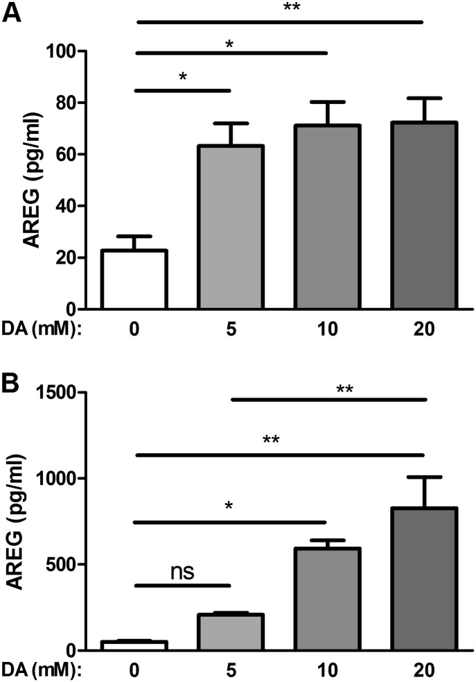
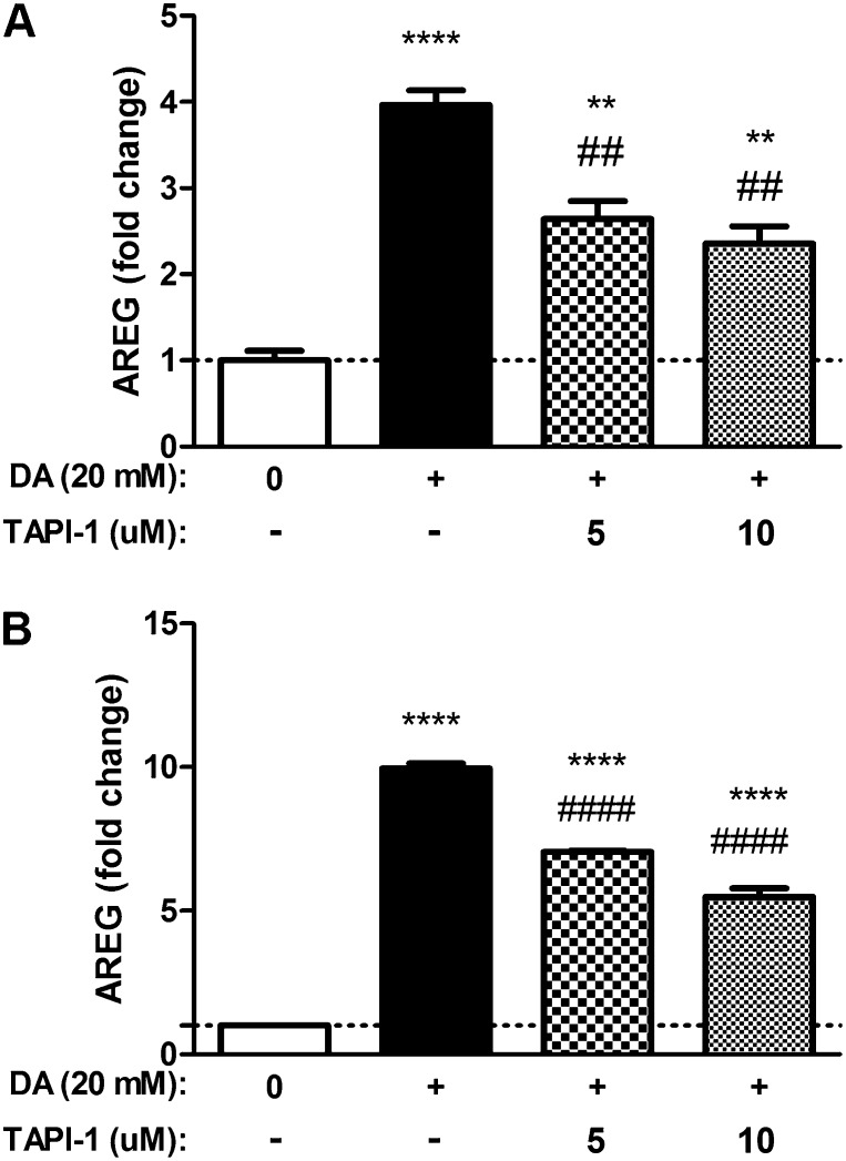
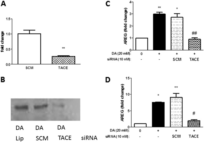
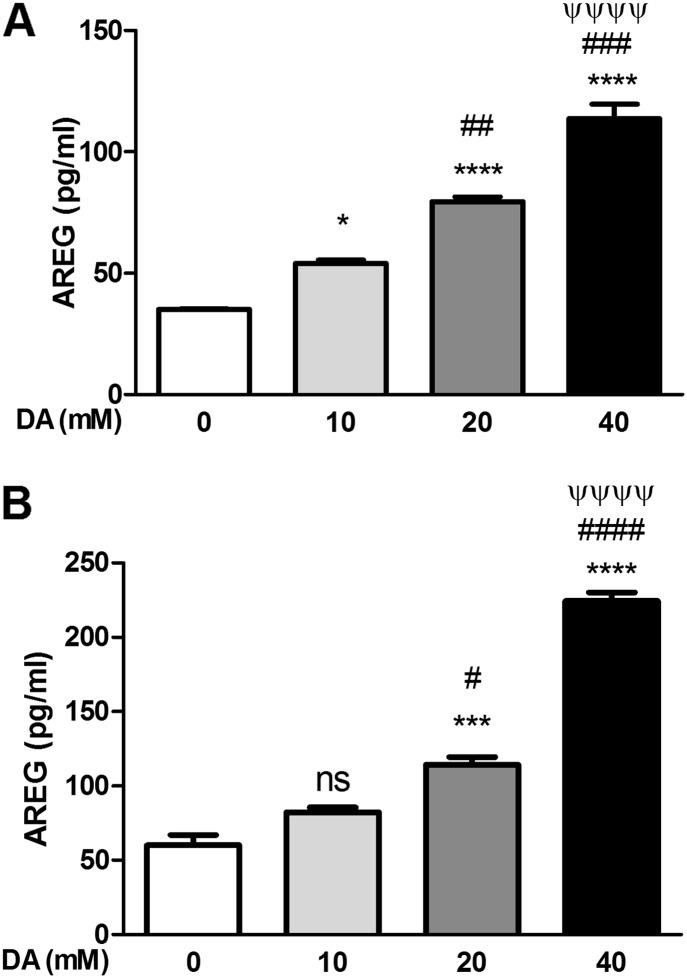
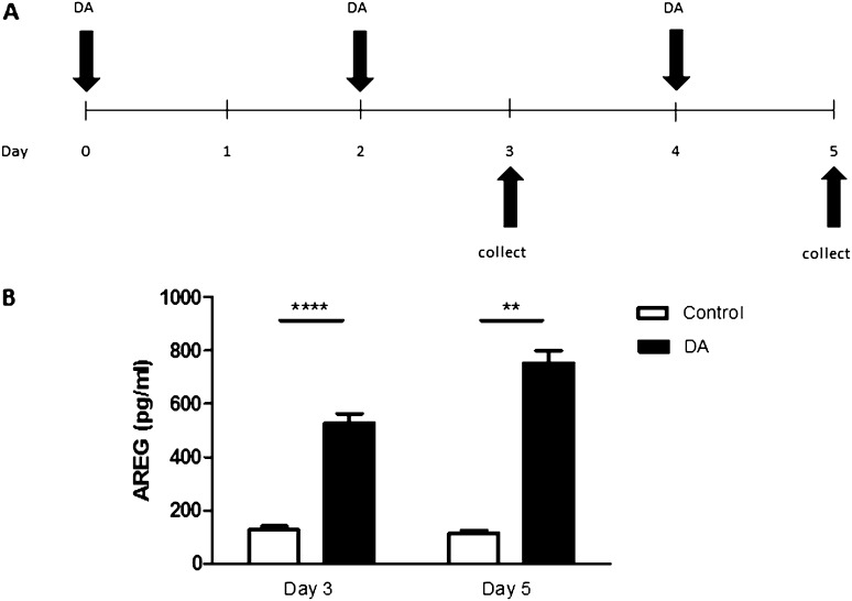
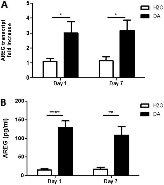
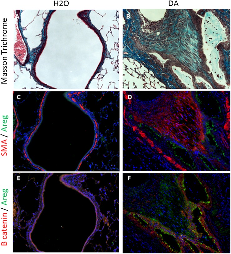
Diacetyl (DA), a component of artificial butter flavoring, has been linked to the development of bronchiolitis obliterans (BO), a disease of airway epithelial injury and airway fibrosis. The epidermal growth factor receptor ligand, amphiregulin (AREG), has been implicated in other types of epithelial injury and lung fibrosis. We investigated the effects of DA directly on the pulmonary epithelium, and we hypothesized that DA exposure would result in epithelial cell shedding of AREG. Consistent with this hypothesis, we demonstrate that DA increases AREG by the pulmonary epithelial cell line NCI-H292 and by multiple independent primary human airway epithelial donors grown under physiologically relevant conditions at the air-liquid interface. Furthermore, we demonstrate that AREG shedding occurs through a TNF-α-converting enzyme (TACE)-dependent mechanism via inhibition of TACE activity in epithelial cells using the small molecule inhibitor, TNF-α protease inhibitor-1, as well as TACE-specific small inhibitor RNA. Finally, we demonstrate supportive in vivo results showing increased AREG transcript and protein levels in the lungs of rodents with DA-induced BO. In summary, our novel in vitro and in vivo observations suggest that further study of AREG is warranted in the pathogenesis of DA-induced BO.

Keywords: TNF-α–converting enzyme; amphiregulin; bronchiolitis obliterans; diacetyl; epidermal growth factor receptor.


Figures

Figure 1.
Figure 1.
Diacetyl (DA) induces amphiregulin (AREG) shedding in human epithelial NCI-H292 cells. Cells were exposed to increasing concentrations of DA for 30 minutes and supernatants were assessed at 6 hours (A) or 24 hours (B). Increased AREG shedding was detected at both 6 and 24 hours. Data shown are averaged across three independent experiments. Data presented are mean (± SD), where DA-exposed groups were compared with Hanks’ balanced salt solution (HBSS) control, by one-way ANOVA with Bonferroni’s correction. **P < 0.01, *P < 0.05. ns, nonsignificant.
Figure 2.
Figure 2.
DA-induced AREG shedding by NCI-H292 epithelial cells is attenuated by TNF-α protease inhibitor (TAPI)-1. Cells were exposed to 20 mM DA for 30 minutes, followed by TAPI-1 treatment (5 or 10 μM) for 6 or 24 hours. AREG shedding was measured at 6 hours (A) and 24 hours (B). Treatment with TAPI-1 reduced AREG shedding at both 6 and 24 hours. Data shown are representative results (n = 3), reproduced over three independent experiments. Data presented are mean (± SD) from a single representative experiment, where DA-exposed groups were compared with HBSS control (*) or TAPI-1–treated DA-exposed samples were compared with DA treated alone (#), by nonparametric t test. ****,####P < 0.0001, **,##P < 0.01.
Figure 3.
Figure 3.
DA-induced AREG shedding by airway epithelial cells is mediated by TNF-α–converting enzyme (TACE). DA-increased AREG shedding by human NCI-H292 epithelial cells is blocked by treatment with TACE small inhibitor (si)RNA. Cells were transfected with siRNA containing a nonspecific scramble sequence (SCM) or a TACE-specific sequence (TACE). Cells transfected with lipofectamine only (Lip) served as a vector control. Knockdown of TACE with TACE-specific siRNA reduced TACE transcript (A) and protein (B) levels at 40 hours. Transfected cells were treated with DA for 30 minutes, and AREG shedding was measured by ELISA in culture medium at 6 hours (C) or 24 hours (D) after exposure. In cells with attenuated levels of TACE, AREG shedding was reduced at both time points. Data presented are mean (± SD), where DA-exposed groups were compared with HBSS control (*) or TACE siRNA–treated DA-exposed samples were compared with DA treated alone (#) by nonparametric t test. *,#P < 0.05, **P < 0.01, ##P < 0.01.
Figure 4.
Figure 4.
DA induces AREG shedding by primary normal human bronchial epithelial (NHBE) cells. Cells were exposed apically with increasing concentrations of DA for 30 minutes. AREG protein levels were measured in basolateral culture medium by ELISA at 6 hours (A) or 24 hours (B). Increased AREG shedding was detected at both 6 and 24 hours. Data shown are representative results (n = 3), reproduced over three independent experiments, with similar results reproduced across multiple donors (Figure E1). Data presented are mean (± SD), where DA-exposed groups were compared with HBSS control (*), the 5-mM DA group was compared with 10 and 20 mM DA (#), and 10 mM compared with 20 mM DA (ψ), using one-way ANOVA with Bonferroni’s correction ****,####,ψψψψP < 0.0001, ***,###P < 0.001, ##P < 0.01, *,#P < 0.05. ns, nonsignificant.
Figure 5.
Figure 5.
DA vapor exposure induces AREG shedding by primary human airway epithelial cells. (A) DA vapor exposure model: EpiAirway tissuesTM, grown at air–liquid interface (ALI), were washed and exposed to DA for 1 hour on Days 0, 2, and 4. Basolateral supernatants were collected and analyzed for AREG on Days 3 and 5. (B) DA vapor induced AREG shedding by primary human airway epithelial cells. Basolateral AREG shedding was measured by ELISA in culture medium 24 hours after the second (Day 3) or third (Day 5) exposures. Data presented are mean (± SD) for two control or three DA-treated tissue inserts derived from a single donor assayed in duplicate. ****P < 0.0001, **P < 0.01, by nonparametric t test.
Figure 6.
Figure 6.
AREG transcript and protein levels are increased in vivo after DA exposure. Whole-lung AREG transcripts (A) and AREG protein in bronchoalveolar lavage fluid (B) were determined at Days 1 and 7 in rats after exposure to vehicle (n = 6) or DA (n = 8). Data presented are mean (± SD). ****P < 0.0001, **P < 0.01, *P < 0.05, by nonparametric t test.
Figure 7.
Figure 7.
Large airway epithelial AREG protein is overexpressed in DA-induced bronchiolitis obliterans (BO). Rodent tissue was assessed for collagen using Masson trichrome staining (A and B), AREG (green) (CF), smooth muscle actin (SMA; red) (C and D), and β-catenin (red) (E and F), and nuclear counterstained with 4′,6-diamidino-2-phenylindole (blue) 7 days after DA exposure. Control airways (A, C, and E) illustrate minimal collagen, AREG, and SMA expression and junctional β-catenin within the airway regions. Fibrotic airways from a DA-treated rodent (B, D, and F) express increased collagen, AREG, and SMA expression within the airway epithelium. Magnification of all images is 200×.

References

    1. Estenne M, Maurer JR, Boehler A, Egan JJ, Frost A, Hertz M, Mallory GB, Snell GI, Yousem S. Bronchiolitis obliterans syndrome 2001: an update of the diagnostic criteria. J Heart Lung Transplant. 2002;21:297–310. - PubMed
    1. Kreiss K, Gomaa A, Kullman G, Fedan K, Simoes EJ, Enright PL. Clinical bronchiolitis obliterans in workers at a microwave-popcorn plant. N Engl J Med. 2002;347:330–338. - PubMed
    1. Hubbs AF, Battelli LA, Goldsmith WT, Porter DW, Frazer D, Friend S, Schwegler-Berry D, Mercer RR, Reynolds JS, Grote A, et al. Necrosis of nasal and airway epithelium in rats inhaling vapors of artificial butter flavoring. Toxicol Appl Pharmacol. 2002;185:128–135. - PubMed
    1. Hubbs AF, Goldsmith WT, Kashon ML, Frazer D, Mercer RR, Battelli LA, Kullman GJ, Schwegler-Berry D, Friend S, Castranova V. Respiratory toxicologic pathology of inhaled diacetyl in Sprague-Dawley rats. Toxicol Pathol. 2008;36:330–344. - PubMed
    1. Palmer SM, Flake GP, Kelly FL, Zhang HL, Nugent JL, Kirby PJ, Foley JF, Gwinn WM, Morgan DL. Severe airway epithelial injury, aberrant repair and bronchiolitis obliterans develops after diacetyl instillation in rats. PLoS ONE. 2011;6:e17644. - PMC - PubMed

Publication types

MeSH terms