. 2014 May 9;9(5):e97414.
doi: 10.1371/journal.pone.0097414. eCollection 2014.

The synergistic in vitro and in vivo antitumor effect of combination therapy with salinomycin and 5-fluorouracil against hepatocellular carcinoma

Affiliations

The synergistic in vitro and in vivo antitumor effect of combination therapy with salinomycin and 5-fluorouracil against hepatocellular carcinoma

Fan Wang et al. PLoS One. .

Abstract

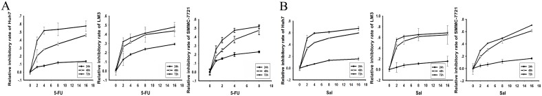
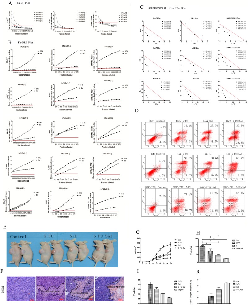
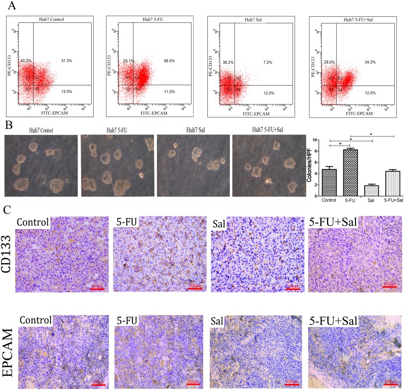
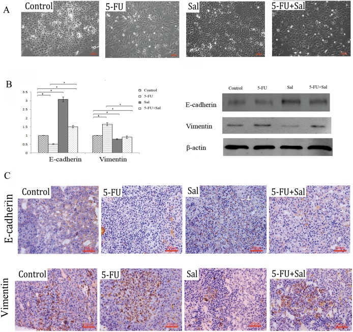
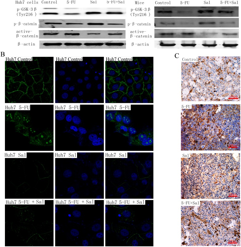
Hepatocellular carcinoma (HCC) is one of the few cancers in which a continuous increase in incidence has been observed over several years. Drug resistance is a major problem in the treatment of HCC. In the present study, we used salinomycin (Sal) and 5-fluorouracil (5-FU) combination therapy on HCC cell lines Huh7, LM3 and SMMC-7721 and nude mice subcutaneously tumor model to study whether Sal could increase the sensitivity of hepatoma cells to the traditional chemotherapeutic agent such as 5-FU. The combination of Sal and 5-FU resulted in a synergistic antitumor effect against liver tumors both in vitro and in vivo. Sal reversed the 5-FU-induced increase in CD133(+) EPCAM(+) cells, epithelial-mesenchymal transition and activation of the Wnt/β-catenin signaling pathway. The combination of Sal and 5-FU may provide us with a new approach to reverse drug resistant for the treatment of patients with HCC.


Conflict of interest statement

Competing Interests: The authors have declared that no competing interests exist.

Figures

Figure 1
Figure 1. Growth inhibition curves for HCC cell lines Huh7, LM3, and SMMC-7721.
5-FU (A) and Sal (B) inhibit HCC cell proliferation. Huh7, LM3, and SMMC-7721 (5×10⁴ cells/ml) were treated with Sal and 5-FU for various times (24, 36, and 48 h). Cell viability was determined using the MTT assay. The data show that Sal and 5-FU exposure reduced Huh7, LM3, and SMMC-7721 cell viability in a dose- and time-dependent manner.
Figure 2
Figure 2. Combination treatment with 5-FU and Sal.
(A–D) Illustrative Fa-CI and Fa-DRI plots for the combination of 5-FU and Sal using different fixed drug ratios. (A) CI values were calculated from each Fa for HCC cell lines Huh7, LM3, and SMMC-7721. Average synergism (CI<1) at Fa>0.5 for all three HCC lines. (B) DRI values were calculated from each Fa for HCC cell lines Huh7, LM3, and SMMC-7721. The 5-FU and Sal chemotherapeutic doses may be significantly reduced (DRI>1) for combinations that are synergistic at Fa>0.5 for all three HCC lines. (C) Isobologram analysis at IC50, IC60 and IC70 for the combinations of HCC cell lines Huh7, LM3, and SMMC-7721. The results indicats synergy, additivity or antagonism when the points are located below, on or above the line, respectively. We can see that (C5-FU, CSal) is located below the line (synergy) at IC 60, IC 70 for HCC cell lines Huh7, LM3 and SMMC-7721. (D) The combination effect of Sal and 5-FU on apoptosis effects were evaluated by flow cytometric analysis. The results showed that combination therapy increased apoptosis of HCC cell lines Huh7, LM3 and SMMC-7721 significantly. (E–R) Combination treatments in the in vivo models (E) Subcutaneous tumor volume following combination therapy was reduced compared to that of the other three groups (two representative mice in each group). (F) HE staining showed the area of apoptosis and necrosis induced by drugs in tumor tissue of treatment group. (G) The tumor growth curve showed that tumor growth rate following combination therapy was slower than that of the other three groups. (H) The relative tumor proliferation rate, VTreatment/VControl, showed that proliferation rate of the combination therapy group was slower than that of the other three groups. (* p <0.05)s. (I) In the combination therapy group, tumor blocks weighed lighter than those of the other three groups (* p <0.05). (R) The tumor growth inhibition rate indicated that the combination therapy significantly inhibited tumor growth than the other three groups (* p <0.05).
Figure 3
Figure 3. Effects of 5-FU, Sal and their combination on cancer stem cell properties in HCC cells.
(A) Flow cytometry assays showed that treatment with 5-FU increased the proportion of the CD133+ EPCAM+ Huh7 cell subpopulation compared with the control group. In contrast, treatment with Sal reduced this proportion compared with the control group. 5-FU combined with Sal reduced this proportion compared with the 5-FU group (*p <0.05). (B) Colony-forming assays were performed to measure the proliferative ability of single cancer cells. The number of colonies increased in the 5-FU treatment group compared with the control group, decreased in the Sal treatment group compared with the control group. The number of colonies was significantly lower in the Sal plus 5-FU combination group compared with the 5-FU treatment group (* p <0.05). (C) Immunohistochemistry indicates CD133+ and EPCAM+ expression in the tumors of mouse xenograft models. (Magnification is 200×).
Figure 4
Figure 4. Effect of 5-FU, Sal, and 5-FU combined with Sal on the epithelial-mesenchymal transition (EMT)-related process.
(A) Morphological changes after the indicated treatment in Huh7 cells (Magnification 200×). (B) Real-time PCR was performed to examine mRNA expression of EMT-related genes (E-cadherin, vimentin) (* p <0.05). Western blot was performed to examine protein expression of EMT-related genes (E-cadherin, vimentin). (C) Immunohistochemistry indicates E-cadherin and vimentin expression in the tumors of mouse xenograft models (Magnification 200×).
Figure 5
Figure 5. Translocation of β-catenin.
(A) The protein expression of p-GSK-3β (Tyr 216) which is active-GSK-3β, p-β-catenin which is inactive β-catenin and active-β-catenin were detected by western-blot in vitro and in vivo. Compared to 5-FU group, p-GSK-3β (Tyr216) expression of Sal group and combination therapy group were significantly up-regulated and we found the similar changes in p-β-catenin protein. Decreased expression of active β-catenin protein were observed in Sal group and combination therapy group compared to 5-FU alone group. (B) Changes in cellular localization of active β-catenin in Huh7 cells. Active β-catenin cellular localization was evaluated by indirect immunofluorescence. Immunofluorescence were labeled of active β-catenin in Huh7 cells (untreated, treated with 5-FU, Sal and Sal plus 5-FU) for 48 h. Nuclei were stained with DAPI, and regions were merged to assess signal colocalization. Magnification is 630×. In the control condition, active β-catenin is present in the cytomembrane and cytoplasm. In the 5-FU treated groups, active β-catenin preferentially accumulates in the nuclear and perinuclear region. In contrast, cells treated with Sal showed preferential localization of active β-catenin in cytomembrane, altering the translocation of active β-catenin to the nucleus. Cells treated with the combination of 5-FU and Sal showed decreased accumulation of β-catenin in the nuclear and perinuclear region compared with the 5-FU treated groups. (C) Immunohistochemistry indicates similar results for β-catenin in the tumors of mouse xenograft models. Magnification is 200×.

Similar articles

Cited by

References

    1. Siegel R, Naishadham D, Jemal A (2013) Cancer statistics, 2013. CA Cancer J Clin 63(1): 11–30. - PubMed
    1. Ikeda M, Okusaka T, Ueno H, Morizane C, Kojima Y, et al. (2008) Predictive factors of outcome and tumor response to systemic chemotherapy in patients with metastatic hepatocellular carcinoma. Jpn J Clin Oncol 38(10): 675–82. - PubMed
    1. Macdonald JS, Astrow AB (2001) Adjuvant therapy of colon cancer. Semin Oncol 28(1): 30–40. - PubMed
    1. Martin M, Pienkowski T, Mackey J, Pawlicki M, Guastalla JP, et al. (2005) Adjuvant docetaxel for node-positive breast cancer. N Engl J Med 352(22): 2302–13. - PubMed
    1. Macdonald JS, Smalley SR, Benedetti J, Hundahl SA, Estes NC, et al. (2001) Chemoradiotherapy after surgery compared with surgery alone for adenocarcinoma of the stomach or gastroesophageal junction. N Engl J Med 345(10): 725–30. - PubMed

MeSH terms