. 2014 May 9;9(5):e97415.
doi: 10.1371/journal.pone.0097415. eCollection 2014.

An optimized triple modality reporter for quantitative in vivo tumor imaging and therapy evaluation

Affiliations

An optimized triple modality reporter for quantitative in vivo tumor imaging and therapy evaluation

Rachel A Levin et al. PLoS One. .

Abstract

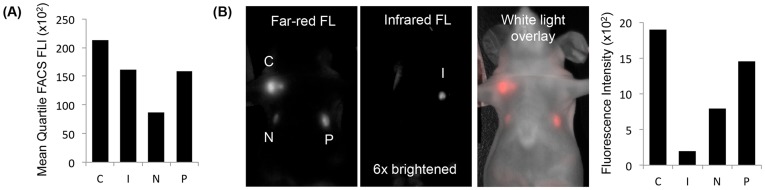
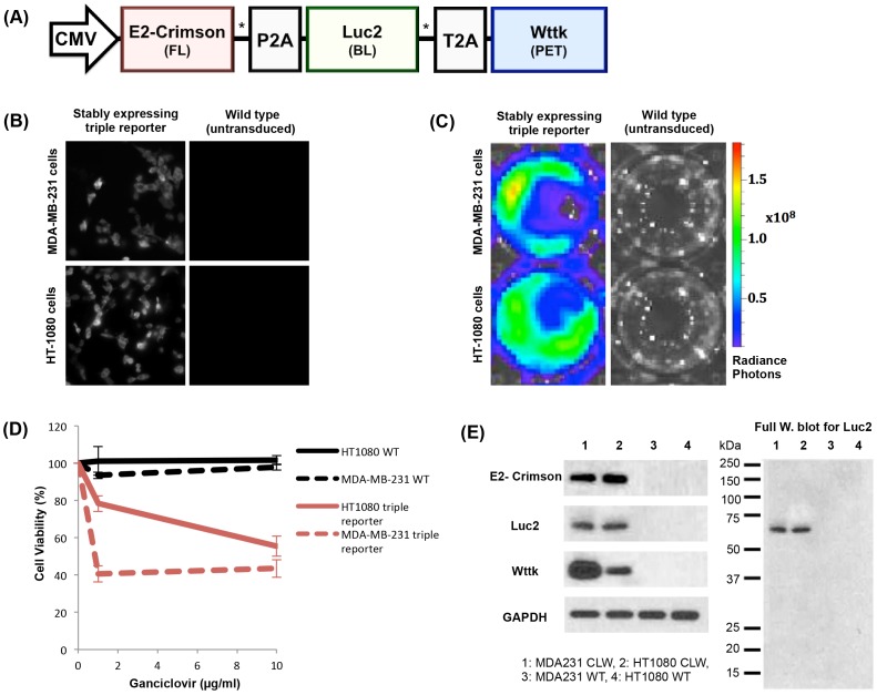
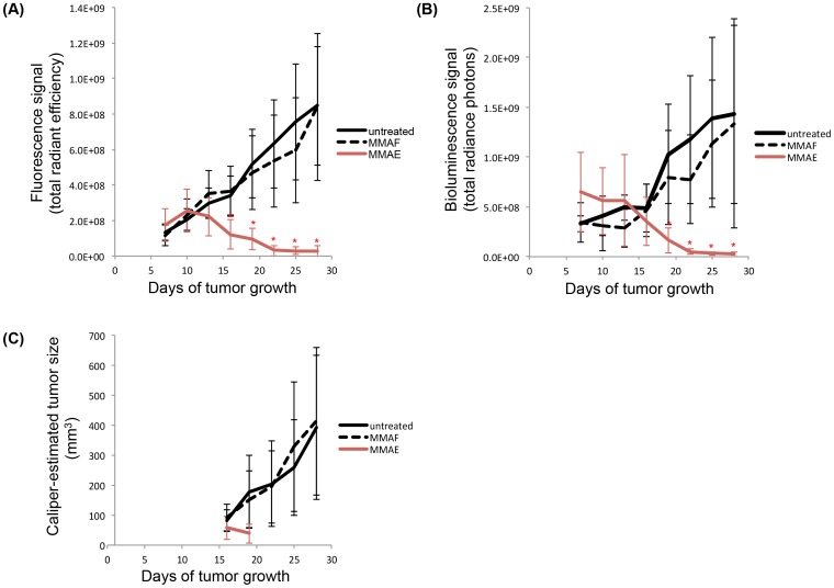
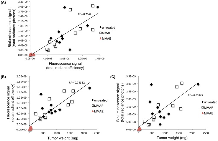
We present an optimized triple modality reporter construct combining a far-red fluorescent protein (E2-Crimson), enhanced firefly luciferase enzyme (Luc2), and truncated wild type herpes simplex virus I thymidine kinase (wttk) that allows for sensitive, long-term tracking of tumor growth in vivo by fluorescence, bioluminescence, and positron emission tomography. Two human cancer cell lines (MDA-MB-231 breast cancer and HT-1080 fibrosarcoma cancer) were successfully transduced to express this triple modality reporter. Fluorescence and bioluminescence imaging of the triple modality reporter were used to accurately quantify the therapeutic responses of MDA-MB-231 tumors to the chemotherapeutic agent monomethyl auristatin E in vivo in athymic nude mice. Positive correlation was observed between the fluorescence and bioluminescence signals, and these signals were also positively correlated with the ex vivo tumor weights. This is the first reported use of both fluorescence and bioluminescence signals from a multi-modality reporter construct to measure drug efficacy in vivo.


Conflict of interest statement

Competing Interests: The authors have declared that no competing interests exist.

Figures

Figure 1
Figure 1. Comparison of four far-red and infrared fluorescent proteins.
(A) The mean quartile (mean 25%) fluorescence intensities (FLI) of E2-Crimson (C), infrared fluorescent protein (I), mNeptune (N), and mPlum (P) in HT-1080 cells measured by fluorescent-activated cell sorting (FACS) were compared (100 mW laser with ex 568 nm and em 650–670 nm for C, N, and P; ex 690 nm and em 710–900 nm for I). (B) The top 5% brightest HT-1080 cells from each fluorescent protein cell type were injected into athymic nude mice (1×10⁶ cells/injection; exposure time: 500 msec; ex 590/23 nm and em 645LP for C, N, and P; ex 640/48 nm and em 700LP for I).
Figure 2
Figure 2. Triple reporter schematic and in vitro validation of components.
(A) The optimized triple modality reporter construct consists of a far-red fluorescent protein (E2-Crimson) for fluorescence imaging (FL), enhanced firefly luciferase enzyme (Luc2) for bioluminescence imaging (BL), and truncated wild type herpes simplex virus I thymidine kinase (wttk) for positron emission tomography (PET). Viral 2A sequences (P2A and T2A) separate each component. A flexible Gly-Ser-Gly linker (*) precedes each 2A sequence. (B) Epifluorescence microscopy of E2-Crimson (580/20 nm excitation filter, 653/95 nm emission filter, 1 second exposure, 40× oil objective) in the wild-type and triple reporter cell lines (stable expression >3 weeks). Images were scaled equally using ImageJ software. (C) Radiance photons (p/s/cm2/sr) produced from Luc2 activity in the triple reporter cell lines (stable expression >3 weeks) compared to their respective wild type cell lines (7.4×10⁴ cells/well) immediately after exposure to D-luciferin (150 µg/ml). The wells shown are representative of triplicate wells in a 48-well plate. (D) Wttk activity measured by cell death after 6 days of ganciclovir treatment for the triple reporter cell lines (stable expression >3 weeks) compared to their respective wild type (WT) cell lines. Only viable cells can convert the CellTiter 96 AQueous One Solution into a product with an absorbance at 490 nm, which was used to calculate the cell viability. (E) Expression of each individual reporter protein in the triple reporter (CLW) cell lines compared to their respective wild type (WT) cell lines shown by Western blot (3×10⁴ cells/well). Successful self-cleavage of viral 2A sequences was observed in all Western blots, as shown in the representative full Western blot for Luc2. A fusion protein consisting of all three modalities (∼135 kDa) or two of the successive modalities (∼95 and ∼106 kDa for E2-Crimson+Luc2 and Luc2+wttk, respectively) was not observed.
Figure 3
Figure 3. In vivo validation of the triple reporter components.
Representative live animal images from the coronal plane of six MDA-MB-231 triple reporter tumors and two HT-1080 triple reporter tumors confirm the activity of all three imaging modalities in both cell lines in vivo. The fluorescence signal is shown as the radiant efficiency (p/s/cm2/str)/(mW/cm2). The bioluminescence signal is shown as the radiance photons (p/s/cm2/sr). The PET signal is shown as the% injected dose of 18F-FHBG per gram. High gut (GI) retention is characteristic of 18F-FHBG in microPET imaging, which required that tumors be placed away from the abdomen of each mouse.
Figure 4
Figure 4. Quantification of the triple reporter optical signals to monitor therapy responses in vivo.
(A) Average fluorescence signal [(p/s)/(cm2/sr)] of each MDA-MB-231 triple reporter tumor treatment group over time. (B) Average bioluminescence signal (p/s) of each MDA-MB-231 triple reporter tumor treatment group over time. (C) Average size (mm3) of each MDA-MB-231 triple reporter tumor treatment group over time based on caliper measurements. MMAE or MMAF (0.5 nmol/g) was administered on days 7, 10, 13, 16, 19, and 22. Significant decreases (p<0.005) in the tumor optical signals in the MMAE-treated group compared to the untreated and MMAF-treated groups are indicated by*.
Figure 5
Figure 5. Correlation between triple reporter tumor optical signals in vivo and weights ex vivo following therapy.
(A) Fluorescence signal [(p/s)/(cm2/sr)] vs. bioluminescence signal (p/s) of the MDA-MB-231 triple reporter tumors on day 28 of tumor growth. (B) Fluorescence signal [(p/s)/(cm2/sr)] vs. weight (mg) of the MDA-MB-231 triple reporter tumors on day 28 of tumor growth. (C) Bioluminescence signal (p/s) vs. weight (mg) of the MDA-MB-231 triple reporter tumors on day 28 of tumor growth.

References

    1. Massoud TF, Gambhir SS (2003) Molecular imaging in living subjects: seeing fundamental biological processes in a new light. Genes Dev 17: 545–580. - PubMed
    1. Ray P, De A, Min JJ, Tsien RY, Gambhir SS (2004) Imaging tri-fusion multimodality reporter gene expression in living subjects. Cancer Res 64: 1323–1330. - PMC - PubMed
    1. Ponomarev V, Doubrovin M, Serganova I, Vider J, Shavrin A, et al. (2004) A novel triple-modality reporter gene for whole-body fluorescent, bioluminescent, and nuclear noninvasive imaging. Eur J Nucl Med Mol Imaging 31: 740–751. - PubMed
    1. Kesarwala AH, Prior JL, Sun J, Harpstrite SE, Sharma V, et al. (2006) Second-generation triple reporter for bioluminescence, micro-positron emission tomography, and fluorescence imaging. Mol Imaging 5: 465–474. - PubMed
    1. Ray P, Tsien R, Gambhir SS (2007) Construction and validation of improved triple fusion reporter gene vectors for molecular imaging of living subjects. Cancer Res 67: 3085–3093. - PubMed

Publication types