

Amygdala perfusion is predicted by its functional connectivity with the ventromedial prefrontal cortex and negative affect
- PMID: 24816735
- PMCID: PMC4016310
- DOI: 10.1371/journal.pone.0097466
Amygdala perfusion is predicted by its functional connectivity with the ventromedial prefrontal cortex and negative affect
Abstract
Background: Previous studies have shown that the activity of the amygdala is elevated in people experiencing clinical and subclinical levels of anxiety and depression (negative affect). It has been proposed that a reduction in inhibitory input to the amygdala from the prefrontal cortex and resultant over-activity of the amygdala underlies this association. Prior studies have found relationships between negative affect and 1) amygdala over-activity and 2) reduced amygdala-prefrontal connectivity. However, it is not known whether elevated amygdala activity is associated with decreased amygdala-prefrontal connectivity during negative affect states.
Methods: Here we used resting-state arterial spin labeling (ASL) and blood oxygenation level dependent (BOLD) functional magnetic resonance imaging (fMRI) in combination to test this model, measuring the activity (regional cerebral blood flow, rCBF) and functional connectivity (correlated fluctuations in the BOLD signal) of one subregion of the amygdala with strong connections with the prefrontal cortex, the basolateral nucleus (BLA), and subsyndromal anxiety levels in 38 healthy subjects.
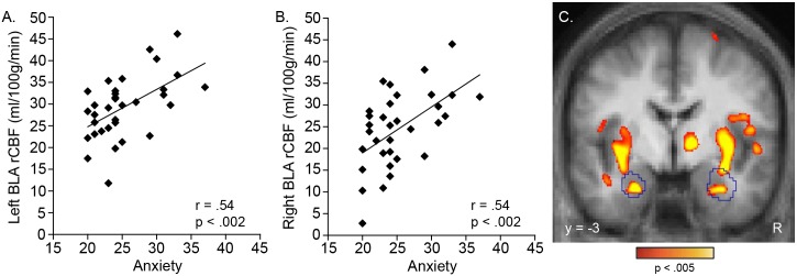
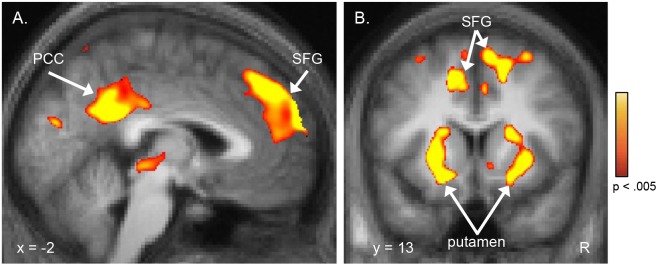
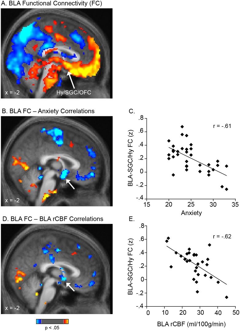
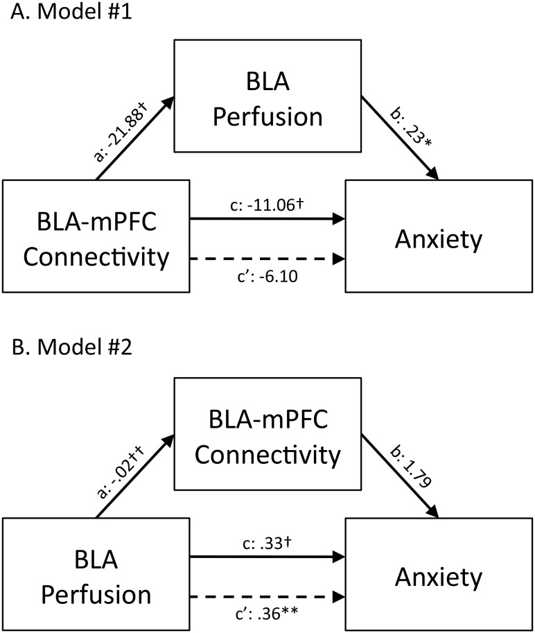
Results: BLA rCBF was strongly correlated with anxiety levels. Moreover, both BLA rCBF and anxiety were inversely correlated with the strength of the functional coupling of the BLA with the caudal ventromedial prefrontal cortex. Lastly, BLA perfusion was found to be a mediator of the relationship between BLA-prefrontal connectivity and anxiety.
Conclusions: These results show that both perfusion of the BLA and a measure of its functional coupling with the prefrontal cortex directly index anxiety levels in healthy subjects, and that low BLA-prefrontal connectivity may lead to increased BLA activity and resulting anxiety. Thus, these data provide key evidence for an often-cited circuitry model of negative affect, using a novel, multi-modal imaging approach.
Conflict of interest statement
Figures
Similar articles
-
Reduced resting-state functional connectivity of the basolateral amygdala to the medial prefrontal cortex in preweaning rats exposed to chronic early-life stress.Brain Struct Funct. 2018 Nov;223(8):3711-3729. doi: 10.1007/s00429-018-1720-3. Epub 2018 Jul 21. Brain Struct Funct. 2018. PMID: 30032360
-
Development of White Matter Microstructure and Intrinsic Functional Connectivity Between the Amygdala and Ventromedial Prefrontal Cortex: Associations With Anxiety and Depression.Biol Psychiatry. 2017 Oct 1;82(7):511-521. doi: 10.1016/j.biopsych.2017.01.008. Epub 2017 Jan 17. Biol Psychiatry. 2017. PMID: 28274468 Free PMC article.
-
Modulating functional connectivity between medial frontopolar cortex and amygdala by inhibitory and excitatory transcranial magnetic stimulation.Hum Brain Mapp. 2019 Oct 15;40(15):4301-4315. doi: 10.1002/hbm.24703. Epub 2019 Jul 3. Hum Brain Mapp. 2019. PMID: 31268615 Free PMC article.
-
Altered corticolimbic connectivity reveals sex-specific adolescent outcomes in a rat model of early life adversity.Elife. 2020 Jan 20;9:e52651. doi: 10.7554/eLife.52651. Elife. 2020. PMID: 31958061 Free PMC article.
-
Anxiety and depression: A top-down, bottom-up model of circuit function.Ann N Y Acad Sci. 2023 Jul;1525(1):70-87. doi: 10.1111/nyas.14997. Epub 2023 May 2. Ann N Y Acad Sci. 2023. PMID: 37129246 Free PMC article. Review.
Cited by
-
Neurogenetic plasticity and sex influence the link between corticolimbic structural connectivity and trait anxiety.Sci Rep. 2017 Sep 8;7(1):10959. doi: 10.1038/s41598-017-11497-2. Sci Rep. 2017. PMID: 28887539 Free PMC article.
-
An Honest Reckoning With the Amygdala and Mental Illness.Am J Psychiatry. 2024 Dec 1;181(12):1059-1075. doi: 10.1176/appi.ajp.20240941. Am J Psychiatry. 2024. PMID: 39616453 Free PMC article. Review.
-
Dopaminergic Mechanisms Underlying Normal Variation in Trait Anxiety.J Neurosci. 2019 Apr 3;39(14):2735-2744. doi: 10.1523/JNEUROSCI.2382-18.2019. Epub 2019 Feb 8. J Neurosci. 2019. PMID: 30737306 Free PMC article.
-
Dysfunctional Limbic Circuitry Underlying Freezing of Gait in Parkinson's Disease.Neuroscience. 2018 Mar 15;374:119-132. doi: 10.1016/j.neuroscience.2018.01.044. Epub 2018 Jan 31. Neuroscience. 2018. PMID: 29408498 Free PMC article.
-
GABA content within the ventromedial prefrontal cortex is related to trait anxiety.Soc Cogn Affect Neurosci. 2016 May;11(5):758-66. doi: 10.1093/scan/nsv155. Epub 2015 Dec 31. Soc Cogn Affect Neurosci. 2016. PMID: 26722018 Free PMC article.
References
-
- Jackson J (1931–1932) In: Taylor J, editor. Selected writings of John Hughlings Jackson. London: Hodder.
-
- Ey H (1978) Hughlings Jackson’s fundamental principles applied to psychiatry. In: R H, editor. Historical Explorations in Medicine and Psychiatry. New York: Springer. 204–219.
-
- Mayberg HS (2003) Modulating dysfunctional limbic-cortical circuits in depression: towards development of brain-based algorithms for diagnosis and optimised treatment. Br Med Bull 65: 193–207. - PubMed
-
- Davidson RJ (2002) Anxiety and affective style: role of prefrontal cortex and amygdala. Biol Psychiatry 51: 68–80. - PubMed
-
- Shin LM, Rauch SL, Pitman RK (2006) Amygdala, medial prefrontal cortex, and hippocampal function in PTSD. Ann N Y Acad Sci 1071: 67–79. - PubMed
Publication types
MeSH terms
Substances
Grants and funding
LinkOut - more resources
Full Text Sources
Other Literature Sources
Medical
