. 2014 May 9;9(5):e96903.
doi: 10.1371/journal.pone.0096903. eCollection 2014.

Fc receptors for immunoglobulins and their appearance during vertebrate evolution

Affiliations

Fc receptors for immunoglobulins and their appearance during vertebrate evolution

Srinivas Akula et al. PLoS One. .

Erratum in

Abstract

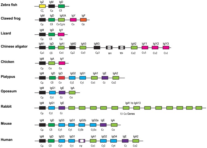
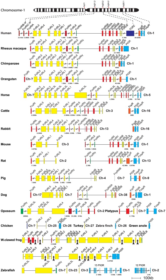
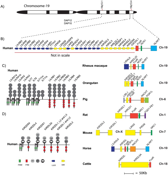
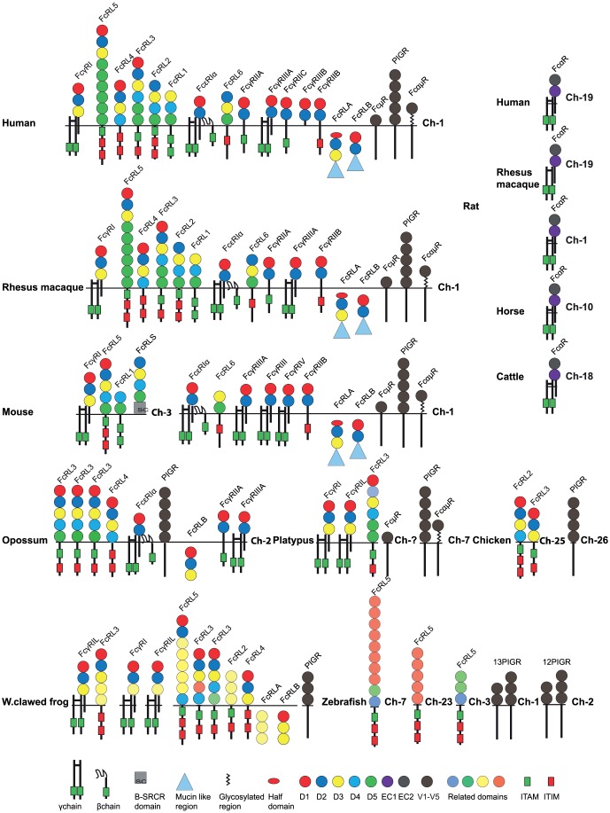
Receptors interacting with the constant domain of immunoglobulins (Igs) have a number of important functions in vertebrates. They facilitate phagocytosis by opsonization, are key components in antibody-dependent cellular cytotoxicity as well as activating cells to release granules. In mammals, four major types of classical Fc receptors (FcRs) for IgG have been identified, one high-affinity receptor for IgE, one for both IgM and IgA, one for IgM and one for IgA. All of these receptors are related in structure and all of them, except the IgA receptor, are found in primates on chromosome 1, indicating that they originate from a common ancestor by successive gene duplications. The number of Ig isotypes has increased gradually during vertebrate evolution and this increase has likely been accompanied by a similar increase in isotype-specific receptors. To test this hypothesis we have performed a detailed bioinformatics analysis of a panel of vertebrate genomes. The first components to appear are the poly-Ig receptors (PIGRs), receptors similar to the classic FcRs in mammals, so called FcRL receptors, and the FcR γ chain. These molecules are not found in cartilagous fish and may first appear within bony fishes, indicating a major step in Fc receptor evolution at the appearance of bony fish. In contrast, the receptor for IgA is only found in placental mammals, indicating a relatively late appearance. The IgM and IgA/M receptors are first observed in the monotremes, exemplified by the platypus, indicating an appearance during early mammalian evolution. Clearly identifiable classical receptors for IgG and IgE are found only in marsupials and placental mammals, but closely related receptors are found in the platypus, indicating a second major step in Fc receptor evolution during early mammalian evolution, involving the appearance of classical IgG and IgE receptors from FcRL molecules and IgM and IgA/M receptors from PIGR.


Conflict of interest statement

Competing Interests: The authors have declared that no competing interests exist.

Figures

Figure 1
Figure 1. The immunoglobulin heavy chain locus of a panel of selected vertebrates from fish to humans.
The locus depicts every single gene as a block and without individual exons. The figure is not to scale and the genes have been color coded; IgM in black, IgD in dark green, IgA and IgX in light green, IgY in magenta, IgG in blue, IgE in purple, IgO in red, IgZ in yellow, IgF in orange and pseudogenes in shaded grey or purple.
Figure 2
Figure 2. The major Fc receptor gene loci in a panel of different vertebrates.
Each horizontal line corresponds to a chromosome on which different Fc receptor genes are located. Genes are color coded. The Fc receptor-like (FcRL) genes are shown in yellow except the FcRLA and B that are in light green, classical IgG receptors in red (pseudogenes in striped red), IgE receptor in dark green, the IgM receptor in orange, the poly-Ig receptor (PIGR) and the IgA/IgM receptor in blue. The figure contains the genes identified for the following animal species: Zebrafish (Danio rerio), Western clawed frog (Xenopus silunarana and Xenopus tropicalis), Chicken (Gallus gallus), Turkey (Meleagris gallopovo), Zebra Finch (Taeniopygia guttata), Green anole (Anolis carolinennsis), Platypus (Ornithorhynchus anatinus), Opossum (Monodelphis domestica), Rat (Ratuus norvegius), Mouse (Mus musculus), Horse (Equus caballus), Rabbit (Oryctologus cunicullus), Pig (Sus scrofa), Dog (Canis lupus familaris), Cattle (Bos taurus), Orangutan (Pongo abelli), Chimpanzee (Pan troglodytes), Rhesus macaque (Macaca mulatta), Human (Homo sapiens).
Figure 3
Figure 3. The FcαR locus and neighboring genes.
Panel A shows a schematic drawing of chromosome 19, panel B a schematic drawing of the LRC locus, panel C the domain structure of the LILRs of this locus (an ‘R’ represents arginine in the transmembrane region) and panel D the domain structure of the KIRs. In the detailed map of the region encoding the IgA receptor each horizontal line corresponds to a chromosome on which different Fc receptor genes are located. Genes are color coded. The IgA receptor in red, the KIRs and LILRs in yellow, the neighboring genes that are used as markers for the chromosomal region, NCR1 in dark green, the NACHT in orange, the NLRP7 in light blue, the PGRL1 in light green and the FQD1-FB1 in dark blue. The overall structure of the LRC locus and the protein domain structures have been adopted from Espeli et al 2010 and Brown et al 2004 , .
Figure 4
Figure 4. A phylogenetic tree of Fc receptor sequences from a panel of vertebrates analyzed with the MrBayes program.
The Maximum-likelihood tree was based on the Bayesian methods of phylogenetic interference. Robustness of nodes was tested with the posterior probabilities based on MCMC analysis as implemented in the MrBayes program. Node supported posterior values are given in the phylogenetic tree. Groups of genes that are more closely related, thereby forming a sub-branch, are indicated by light or dark grey shading to make them more easily visible.
Figure 5
Figure 5. A summary of domain structures and signaling motifs of the various vertebrate Fc receptors.
The Ig-like domains are depicted as filled circles with color-coding according to the similarities in sequence based on phylogenetic analyses , . The domain type D1, D2, D3, D4 and D5 show a relatively conserved pattern in most tetrapods and have therefore been color-coded in red dark blue, yellow, light blue and green. A phylogenetic analysis of all the individual domains presented in figure 5 and a few additional receptors are presented in supplementary figure S4. The color-coding in figure 5 is based on this supplementary figure. The extracellular regions, the transmembrane regions and cytoplasmic tails are not to scale in order to show the positions of potential signaling motifs like ITAMs (green boxes) and ITIMs (red boxes), which regulate the biological function the Fc receptors. Non-consensus ITIMs are also indicated as boxes with half red half white. Some of the intracellular proteins contain C-terminal mucin-like regions that are depicted as blue triangles.

References

    1. Zhao Y, Cui H, Whittington CM, Wei Z, Zhang X, et al. (2009) Ornithorhynchus anatinus (platypus) links the evolution of immunoglobulin genes in eutherian mammals and nonmammalian tetrapods. J Immunol 183: 3285–3293. - PubMed
    1. Zhang YA, Salinas I, Li J, Parra D, Bjork S, et al. (2010) IgT, a primitive immunoglobulin class specialized in mucosal immunity. Nat Immunol 11: 827–835. - PMC - PubMed
    1. Dooley H, Flajnik MF (2006) Antibody repertoire development in cartilaginous fish. Dev Comp Immunol 30: 43–56. - PubMed
    1. Zhang C, Du Pasquier L, Hsu E (2013) Shark IgW C Region Diversification through RNA Processing and Isotype Switching. J Immunol 191: 3410–3418. - PMC - PubMed
    1. Hansen JD, Landis ED, Phillips RB (2005) Discovery of a unique Ig heavy-chain isotype (IgT) in rainbow trout: Implications for a distinctive B cell developmental pathway in teleost fish. Proc Natl Acad Sci U S A 102: 6919–6924. - PMC - PubMed

Publication types

LinkOut - more resources