. 2014 May 9;9(5):e96835.
doi: 10.1371/journal.pone.0096835. eCollection 2014.

Novel polyglutamine model uncouples proteotoxicity from aging

Affiliations

Novel polyglutamine model uncouples proteotoxicity from aging

Nakeirah T M Christie et al. PLoS One. .

Abstract

Polyglutamine expansions in certain proteins are the genetic determinants for nine distinct progressive neurodegenerative disorders and resultant age-related dementia. In these cases, neurodegeneration is due to the aggregation propensity and resultant toxic properties of the polyglutamine-containing proteins. We are interested in elucidating the underlying mechanisms of toxicity of the protein ataxin-3, in which a polyglutamine expansion is the genetic determinant for Machado-Joseph Disease (MJD), also referred to as spinocerebellar ataxia 3 (SCA3). To this end, we have developed a novel model for ataxin-3 protein aggregation, by expressing a disease-related polyglutamine-containing fragment of ataxin-3 in the genetically tractable body wall muscle cells of the model system C. elegans. Here, we demonstrate that this ataxin-3 fragment aggregates in a polyQ length-dependent manner in C. elegans muscle cells and that this aggregation is associated with cellular dysfunction. However, surprisingly, this aggregation and resultant toxicity was not influenced by aging. This is in contrast to polyglutamine peptides alone whose aggregation/toxicity is highly dependent on age. Thus, the data presented here not only describe a new polyglutamine model, but also suggest that protein context likely influences the cellular interactions of the polyglutamine-containing protein and thereby modulates its toxic properties.


Conflict of interest statement

Competing Interests: The authors have declared that no competing interests exist.

Figures

Figure 1
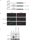
Figure 1. Expression of polyQ-expanded AT3CT in C. elegans body wall muscle cells.
A. Schematic representations of gene constructs. The polyQ-containing C-terminal domain (lacking the N-terminal 257 amino acids) of AT3 was fused to YFP and expressed in body wall muscle cells under the control of the unc-54 promoter to generate Punc-54257cAT3(Qn)::YFP, herein referred to as AT3CT(Qn) B. Fluorescent micrographs showing fixed N2 (wild type), AT3CT(Q45), and AT3CT(Q63) animals imaged for YFP fluorescence (green) or phalloidin-stained actin filaments (red). C. Western blot with an anti-GFP antibody showing relative protein levels for C. elegans expressing YFP alone or AT3CT(Q45) or AT3CT(Q63). The asterisk (*) represents protein running as a GFP monomer. In the AT3CT (Q45) and AT3CT (Q63) lanes, these bands seem to represent cleavage fragments that are likely artifacts of protein extraction.
Figure 2
Figure 2. Aggregation of AT3CT in C. elegans body wall muscle cells.
A. Fluorescence Recovery after Photobleaching (FRAP) of diffuse fluorescent protein in YFP or AT3CT(Q45)-expressing animals, or fluorescent foci in AT3CT(Q45) or AT3(Q63)-expressing animals. Time of bleach is indicated with an arrow. Fluorescence was monitored for 60 s. B. Native gel showing the YFP-containing protein species that accumulate in lines expressing YFP alone, AT3CT(Q45), or AT3CT(Q63). Presumptive aggregates (a), oligomers (o), and monomers (m) are indicated. Fluorescent protein species were visualized under UV light.
Figure 3
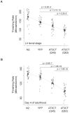
Figure 3. The toxic effects of AT3CT expression in C. elegans body wall muscle cells.
Motility was determined as a measure of thrashing rate in liquid for L4 larvae (A) or animals at day 4 of adulthood (B). All 30 individual data points are represented on a dot blot. The symbol “▪” represents means. 95 percent confidence intervals are indicated. P-values are the results of pairwise student t-test.
Figure 4
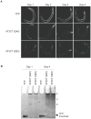
Figure 4. Effect of aging on AT3CT aggregation.
A. Micrographs showing YFP fluorescence in YFP- or AT3CT(Q45 or Q63)-expressing animals. B. Native gels showing the relative distribution of presumptive monomers (m), oligomers (o), and aggregates (a) at day 1 as compared to day 4 of adulthood.
Figure 5
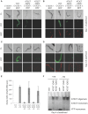
Figure 5. Effect of Heat Shock on C. elegans expressing AT3CT in body wall muscle cells.
Animals expressing a reporter for the heat shock response (PC12C8.1mCherry) or co-expressing this reporter along with YFP, AT3CT(Q45), or AT3CT(Q63) were imaged at day 1 of adulthood before (A) or after (B) a 34°C/15 min heat shock, or at day 4 of adulthood before (C) or after (D) a 34°C/15 min heat shock. E) qRT-PCR showing the relative expression levels of the endogenous C12C8.1 mRNA before and after heat shock. F) Native gel showing in-gel fluorescence from samples taken from YFP, AT3CT(Q45), or AT3CT(Q63) animals after heat shock (+HS) or without heat shock (-HS) at day 1 of adulthood.

Similar articles

Cited by

References

    1. Masino L, Musi V, Menon RP, Fusi P, Kelly G, et al. (2003) Domain architecture of the polyglutamine protein ataxin-3: a globular domain followed by a flexible tail. FEBS Lett 549: 21–25. - PubMed
    1. Ellisdon AM, Thomas B, Bottomley SP (2006) The two-stage pathway of ataxin-3 fibrillogenesis involves a polyglutamine-independent step. J Biol Chem 281: 16888–16896. - PubMed
    1. Chow MK, Ellisdon AM, Cabrita LD, Bottomley SP (2004) Polyglutamine expansion in ataxin-3 does not affect protein stability: implications for misfolding and disease. J Biol Chem 279: 47643–47651. - PubMed
    1. Paulson HL, Perez MK, Trottier Y, Trojanowski JQ, Subramony SH, et al. (1997) Intranuclear inclusions of expanded polyglutamine protein in spinocerebellar ataxia type 3. Neuron 19: 333–344. - PubMed
    1. McLeod CJ, O'Keefe LV, Richards RI (2005) The pathogenic agent in Drosophila models of ‘polyglutamine’ diseases. Hum Mol Genet 14: 1041–1048. - PubMed

Publication types

MeSH terms