

Hard-diet feeding recovers neurogenesis in the subventricular zone and olfactory functions of mice impaired by soft-diet feeding
- PMID: 24817277
- PMCID: PMC4016307
- DOI: 10.1371/journal.pone.0097309
Hard-diet feeding recovers neurogenesis in the subventricular zone and olfactory functions of mice impaired by soft-diet feeding
Abstract
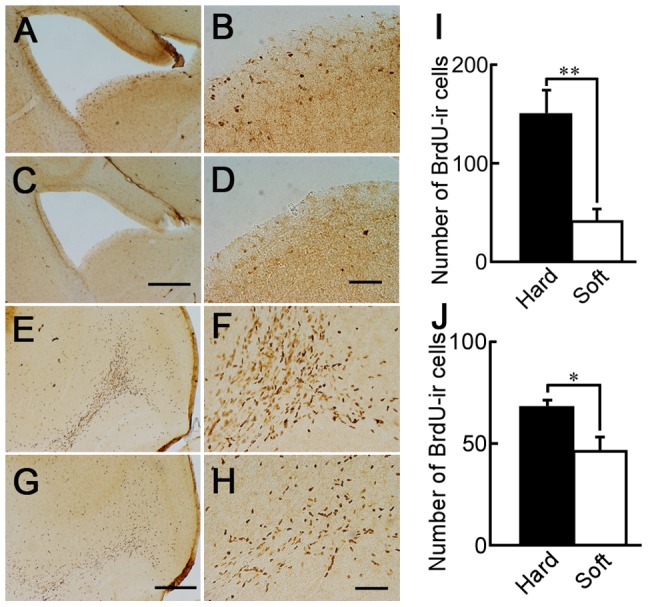
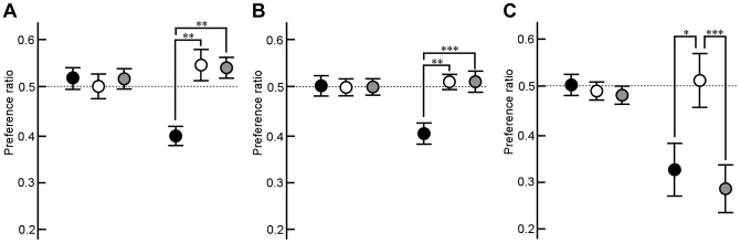
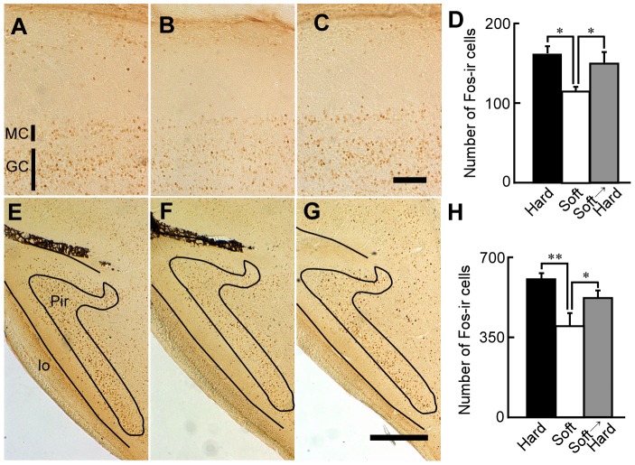
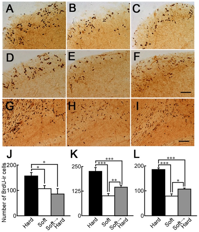
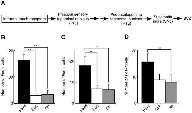
The subventricular zone (SVZ) generates an immense number of neurons even during adulthood. These neurons migrate to the olfactory bulb (OB) and differentiate into granule cells and periglomerular cells. The information broadcast by general odorants is received by the olfactory sensory neurons and transmitted to the OB. Recent studies have shown that a reduction of mastication impairs both neurogenesis in the hippocampus and brain functions. To examine these effects, we first measured the difference in Fos-immunoreactivity (Fos-ir) at the principal sensory trigeminal nucleus (Pr5), which receives intraoral touch information via the trigeminal nerve, when female adult mice ingested a hard or soft diet to explore whether soft-diet feeding could mimic impaired mastication. Ingestion of a hard diet induced greater expression of Fos-ir cells at the Pr5 than did a soft diet or no diet. Bromodeoxyuridine-immunoreactive (BrdU-ir) structures in sagittal sections of the SVZ and in the OB of mice fed a soft or hard diet were studied to explore the effects of changes in mastication on newly generated neurons. After 1 month, the density of BrdU-ir cells in the SVZ and OB was lower in the soft-diet-fed mice than in the hard-diet-fed mice. The odor preferences of individual female mice to butyric acid were tested in a Y-maze apparatus. Avoidance of butyric acid was reduced by the soft-diet feeding. We then explored the effects of the hard-diet feeding on olfactory functions and neurogenesis in the SVZ of mice impaired by soft-diet feeding. At 3 months of hard-diet feeding, avoidance of butyric acid was reversed and responses to odors and neurogenesis were recovered in the SVZ. The present results suggest that feeding with a hard diet improves neurogenesis in the SVZ, which in turn enhances olfactory function at the OB.
Conflict of interest statement
Figures
Similar articles
-
Impaired mastication reduced newly generated neurons at the accessory olfactory bulb and pheromonal responses in mice.Arch Oral Biol. 2014 Dec;59(12):1272-8. doi: 10.1016/j.archoralbio.2014.07.018. Epub 2014 Aug 2. Arch Oral Biol. 2014. PMID: 25150532
-
Soft-diet feeding impairs neural transmission between mitral cells and interneurons in the mouse olfactory bulb.Arch Oral Biol. 2017 Nov;83:209-213. doi: 10.1016/j.archoralbio.2017.07.015. Epub 2017 Jul 29. Arch Oral Biol. 2017. PMID: 28802192
-
Enriched odor exposure increases the number of newborn neurons in the adult olfactory bulb and improves odor memory.J Neurosci. 2002 Apr 1;22(7):2679-89. doi: 10.1523/JNEUROSCI.22-07-02679.2002. J Neurosci. 2002. PMID: 11923433 Free PMC article.
-
Olfactory bulb neurogenesis depending on signaling in the subventricular zone.Cereb Cortex. 2023 Nov 4;33(22):11102-11111. doi: 10.1093/cercor/bhad349. Cereb Cortex. 2023. PMID: 37746807 Review.
-
Is adult neurogenesis essential for olfaction?Trends Neurosci. 2011 Jan;34(1):20-30. doi: 10.1016/j.tins.2010.09.006. Epub 2010 Oct 25. Trends Neurosci. 2011. PMID: 20980064 Review.
Cited by
-
Taste Impairments in a Parkinson's Disease Model Featuring Intranasal Rotenone Administration in Mice.J Parkinsons Dis. 2022;12(6):1863-1880. doi: 10.3233/JPD-223273. J Parkinsons Dis. 2022. PMID: 35848036 Free PMC article.
-
Difference of brain-derived neurotrophic factor expression and pyramid cell count during mastication of food with varying hardness.J Appl Oral Sci. 2019 Apr 1;27:e20180182. doi: 10.1590/1678-7757-2018-0182. J Appl Oral Sci. 2019. PMID: 30970112 Free PMC article.
-
Revisiting the link between cognitive decline and masticatory dysfunction.BMC Geriatr. 2018 Jan 5;18(1):5. doi: 10.1186/s12877-017-0693-z. BMC Geriatr. 2018. PMID: 29304748 Free PMC article. Review.
-
Factors that influence adult neurogenesis as potential therapy.Transl Neurodegener. 2018 Feb 21;7:4. doi: 10.1186/s40035-018-0109-9. eCollection 2018. Transl Neurodegener. 2018. PMID: 29484176 Free PMC article. Review.
-
Short high fat diet triggers reversible and region specific effects in DCX+ hippocampal immature neurons of adolescent male mice.Sci Rep. 2021 Nov 2;11(1):21499. doi: 10.1038/s41598-021-01059-y. Sci Rep. 2021. PMID: 34728755 Free PMC article.
References
-
- Weijenberg RA, Scherder EJ, Lobbezoo F (2011) Mastication for the mind––the relationship between mastication and cognition in ageing and dementia. Neurosci Biobehav Rev 35: 483–497. - PubMed
-
- Lexomboon D, Trulsson M, Wardh I, Parker MG (2012) Chewing ability and tooth loss: association with cognitive impairment in an elderly population study. J Am Geriatr Soc 60: 1951–1956. - PubMed
-
- Stein PS, Desrosiers M, Donegan SJ, Yepes JF, Kryscio RJ (2007) Tooth loss, dementia and neuropathology in the Nun study. J Am Dent Assoc 138: 1314–1322. - PubMed
-
- Miura H, Yamasaki K, Kariyasu M, Miura K, Sumi Y (2003) Relationship between cognitive function and mastication in elderly females. J Oral Rehabil 30: 808–811. - PubMed
-
- Kato T, Usami T, Noda Y, Hasegawa M, Ueda M, et al. (1997) The effect of the loss of molar teeth on spatial memory and acetylcholine release from the parietal cortex in aged rats. Behav Brain Res 83: 239–242. - PubMed
Publication types
MeSH terms
Substances
LinkOut - more resources
Full Text Sources
Other Literature Sources
Miscellaneous
