. 2014 Aug;22(7-8):316-25.
doi: 10.1007/s12471-014-0563-7.

Arrhythmogenic cardiomyopathy: diagnosis, genetic background, and risk management

Affiliations

Arrhythmogenic cardiomyopathy: diagnosis, genetic background, and risk management

J A Groeneweg et al. Neth Heart J. 2014 Aug.

Abstract

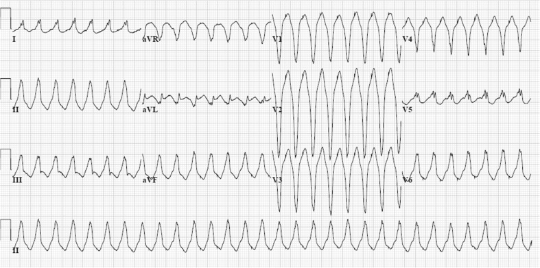
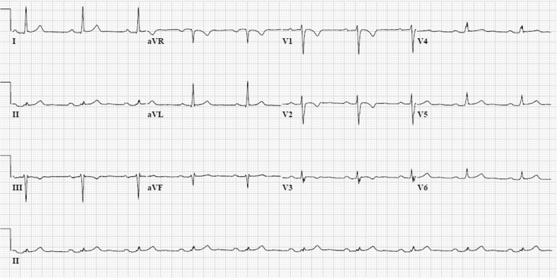
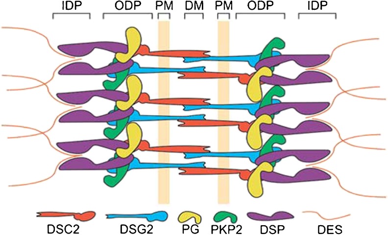
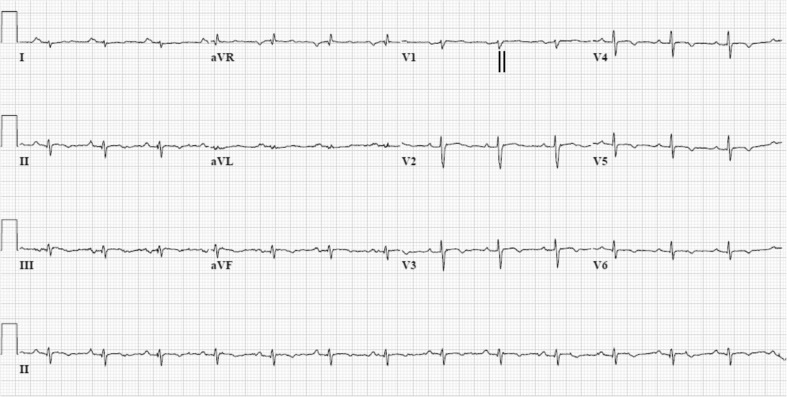
Arrhythmogenic cardiomyopathy (AC), also known as arrhythmogenic right ventricular dysplasia/cardiomyopathy (ARVD/C), is a hereditary disease characterised by ventricular arrhythmias, right ventricular and/or left ventricular dysfunction, and fibrofatty replacement of cardiomyocytes. Patients with AC typically present between the second and the fourth decade of life with ventricular tachycardias. However, sudden cardiac death (SCD) may be the first manifestation, often at young age in the concealed stage of disease. AC is diagnosed by a set of clinically applicable criteria defined by an international Task Force. The current Task Force Criteria are the essential standard for a correct diagnosis in individuals suspected of AC. The genetic substrate for AC is predominantly identified in genes encoding desmosomal proteins. In a minority of patients a non-desmosomal mutation predisposes to the phenotype. Risk stratification in AC is imperfect at present. Genotype-phenotype correlation analysis may provide more insight into risk profiles of index patients and family members. In addition to symptomatic treatment, prevention of SCD is the most important therapeutic goal in AC. Therapeutic options in symptomatic patients include antiarrhythmic drugs, catheter ablation, and ICD implantation. Furthermore, patients with AC and also all pathogenic mutation carriers should be advised against practising competitive and endurance sports.


Figures

Fig. 1
Fig. 1
12-lead ECG of a ventricular tachycardia (VT) in an arrhythmogenic cardiomyopathy (AC) index patient with a pathogenic and a most likely pathogenic plakophilin-2 mutation (c.397C>T p.Gln133* and c.2615C>T p.Thr872Ile). The VT has a left bundle branch block morphology, with inferior axis. The QRS complex is predominantly negative in lead aVL and most positive in lead II, suggesting an origin from the right ventricular outflow tract area in the right ventricle
Fig. 2
Fig. 2
12-lead ECG (during atenolol 25 mg once daily) of the same index patient as in Figure 1. The ECG shows sinus rhythm, horizontal axis, and typical negative T waves in the right precordial leads V1-3. The terminal activation duration (from the nadir of the S wave to the end of all depolarisation deflections) in leads V1-3 is normal (≤55 ms)
Fig. 3
Fig. 3
Schematic representation of the molecular organisation of cardiac desmosomes. The plasma membrane (PM) spanning proteins desmocollin-2 (DSC2) and desmoglein-2 (DSG2) interact in the extracellular space at the dense midline (DM). At the cytoplasmic side, they interact with plakoglobin (PG) and plakophilin-2 (PKP2) at the outer dense plaque (ODP). The PKP2 and PG also interact with desmoplakin (DSP). At the inner dense plaque (IDP), the C-terminus of DSP anchors the intermediate filament desmin (DES). (Source: Reprint with permission from: Van Tintelen et al. Curr Opin Cardiol. 2007;22:185–92)
Fig. 4
Fig. 4
12-lead ECG (while off medications) of a phospholamban founder mutation carrier (c.40_42delAGA, p.Arg14del). The ECG shows sinus rhythm with right axis deviation, low voltages (<0.5 mV in standard leads), and characteristic negative T waves in left precordial leads from V3-6. The terminal activation duration (from the nadir of the S wave to the end of all depolarisation deflections) is 60 ms and therefore prolonged in lead V1 (vertical black lines)

References

    1. Marcus FI, Fontaine GH, Guiraudon G, et al. Right ventricular dysplasia: a report of 24 adult cases. Circulation. 1982;65(2):384–98. doi: 10.1161/01.CIR.65.2.384. - DOI - PubMed
    1. Corrado D, Basso C, Thiene G, et al. Spectrum of clinicopathologic manifestations of arrhythmogenic right ventricular cardiomyopathy/dysplasia: a multicenter study. J Am Coll Cardiol. 1997;30(6):1512–20. doi: 10.1016/S0735-1097(97)00332-X. - DOI - PubMed
    1. Basso C, Thiene G, Corrado D, et al. Arrhythmogenic right ventricular cardiomyopathy. Dysplasia, dystrophy, or myocarditis? Circulation. 1996;94(5):983–91. doi: 10.1161/01.CIR.94.5.983. - DOI - PubMed
    1. Sen-Chowdhry S, Syrris P, Prasad SK, et al. Left-dominant arrhythmogenic cardiomyopathy: an under-recognized clinical entity. J Am Coll Cardiol. 2008;52(25):2175–87. doi: 10.1016/j.jacc.2008.09.019. - DOI - PubMed
    1. Asimaki A, Tandri H, Huang H, et al. A new diagnostic test for arrhythmogenic right ventricular cardiomyopathy. N Engl J Med. 2009;360(11):1075–84. doi: 10.1056/NEJMoa0808138. - DOI - PubMed

LinkOut - more resources