

doi: 10.1016/b978-008045046-9.01046-9.
Olfactory Neuron Patterning and Specification
Affiliations
- PMID: 24817923
- PMCID: PMC4015351
- DOI: 10.1016/b978-008045046-9.01046-9
Item in Clipboard
Olfactory Neuron Patterning and Specification
Dev Neurobiol.
2009.
No abstract available
Figures
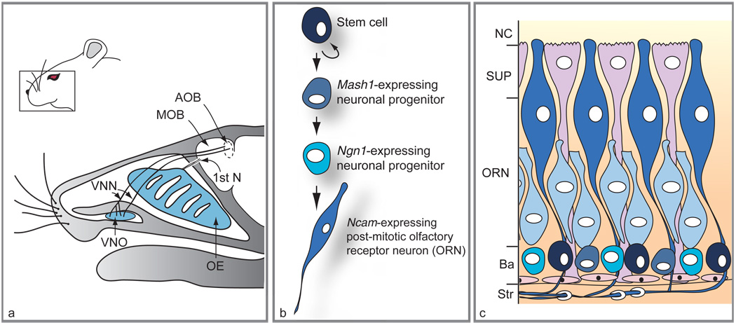
Location of main olfactory epithelium (OE) and vomeronasal organ (VNO) in mouse head, and lineage and distribution of different cell types within the OE. (a) Sagittal section through an adult head reveals the position of the VNO relative to the OE. While the vomeronasal nerves (VNN) target the accessory olfactory bulb (AOB), axons of olfactory receptor neurons (first cranial nerve; 1st N) target the main olfactory bulb (MOB). (b) Scheme of the neuronal differentiation pathway in the OE. (c) Histological arrangement of cells in mature OE. Neuronal stem cells (black) located at the basal layer (Ba) adjacent to the stroma (Str) give rise to transit-amplifying progenitors expressing the Mash1 gene (round gray-blue cells) followed by Ngn1-expressing precursors (turquoise cells). The immature ORNs (light blue elongated cells) arise from the Ngn1-positive cells and mature into Ncam-expressing neurons (dark blue elongated cells). Supporting cells (SUP) lie in a single layer on the apical surface of the OE, at the nasal cavity (NC), while olfactory ensheathing cells wrap around nerve bundles in the underlying stroma. The VNO (not shown) has a similar histological arrangement, with basal cells at the boundary between epithelium and mesenchyme and neurons populating the bulk of the epithelium. Adapted from Kawauchi S, Beites CL, Crocker CE, et al. (2004) Molecular signals regulating proliferation of stem and progenitor cells in mouse olfactory epithelium. Developmental Neuroscience 26: 166–180, with permission. Copyright 2004 by S. Karger AG, Basel, Switzerland.
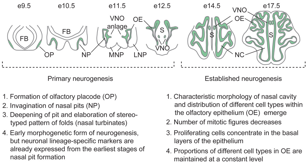
Schematic model and description of primary and established neurogenesis during mouse olfactory epithelium development. Coronal sections through heads at days 9.5–12.5 of gestation (e9.5–e12.5) are depicted for the phase of primary neurogenesis, while horizontal sections at e14.5 and e17.5 (immediately before birth) are portrayed for the phase of established neurogenesis. FB, forebrain; MNP, medial nasal process; VNO, vomeronasal organ; LNP, lateral nasal process; S, nasal septum; NC, nasal cavity. Adapted from Kawauchi S, Shou J, Santos R, et al. (2005) Fgf8 expression defines a morphogenetic center required for olfactory neurogenesis and nasal cavity development in the mouse. Development 132(23): 5211–5223.
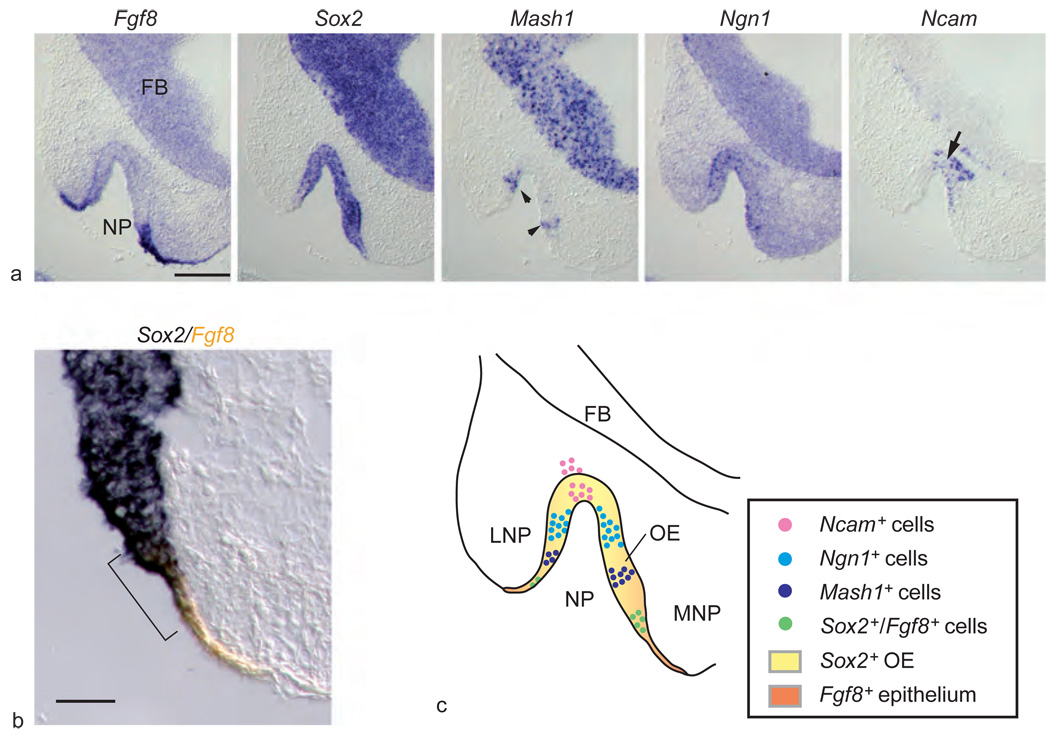
Expression of Fgf8 and neuronal cell markers in developing olfactory epithelium (OE). (a) Five successive images show in situ hybridization for Fgf8 and OE neuronal lineage markers in invaginating nasal pit (NP) at day 10.5 of gestation (FB, forebrain). While Sox2 is expressed throughout the neuroepithelium, Fgf8 is localized to the borders of the invaginating pit. Mash1 (arrowheads) expression is located next to the Fgf8-expressing cells at the inner rim of the nasal pit, while Ncam-expressing neurons (arrow) are located at the center of the pit. (b) Double-label in situ hybridization for Fgf8 (orange) and Sox2 (blue) demonstrates overlap of the two markers in a small rim of surface ectoderm and adjacent invaginating neuroepithelium (bracket). (c) Model of peripheral-to-central process of neuronal differentiation in developing OE and origin of Sox2-expressing neural stem cells from Fgf8-expressing ectoderm (LNP, lateral nasal process; MNP, medial nasal process). Scale bar = 200 µm (a), 50 µm (b). Adapted from Kawauchi S, Shou J, Santos R, et al. (2005) Fgf8 expression defines a morphogenetic center required for olfactory neurogenesis and nasal cavity development in the mouse. Development 132(23): 5211–5223.
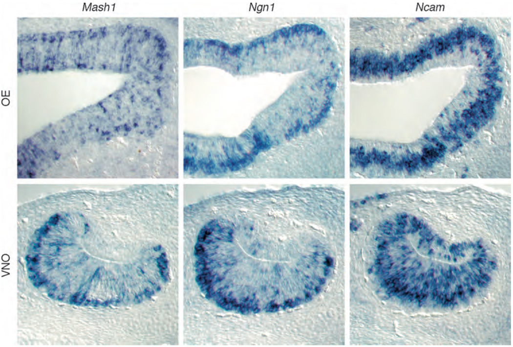
Cell-type-specific markers for committed neuronal progenitors and differentiated olfactory receptor neurons in the established phase of neurogenesis in main olfactory epithelium (OE) and vomeronasal organ (VNO). In situ hybridization is shown for mRNAs encoding Mash1, Ngn1, and Ncam in the OE and VNO of mice at day 14.5 of gestation, when the pattern of neurogenesis becomes established in the two sensory epithelia. Even at this stage in prenatal development, the characteristic laminar patterns of cells are apparent, and the relative proportions of cells at different stages of differentiation are similar to what is seen in the postnatal epithelia. (Top panel) Mash1 and Ngn1 messages are all expressed in basal areas of the OE, while some apical cells express Mash1 but not Ngn1. Ncam, an olfactory receptor neuron marker, is expressed throughout the OE but is absent from the supporting cell layer that lines the nasal cavity. (Bottom panel) Mash1 and Ngn1 are similarly located in the VNO, at the boundary between basal epithelium and underlying connective tissue. Ncam-positive cells populate the majority of the sensory epithelium, from the concave side of the lumen to the basal layer. Adapted from Murray RC, Navi D, Fesenko J, et al. (2003) Widespread defects in the primary olfactory pathway caused by loss of Mash1 function. Journal of Neuroscience 23(5): 1769–1780, with permission. Copyright 2003 by the Society for Neuroscience.
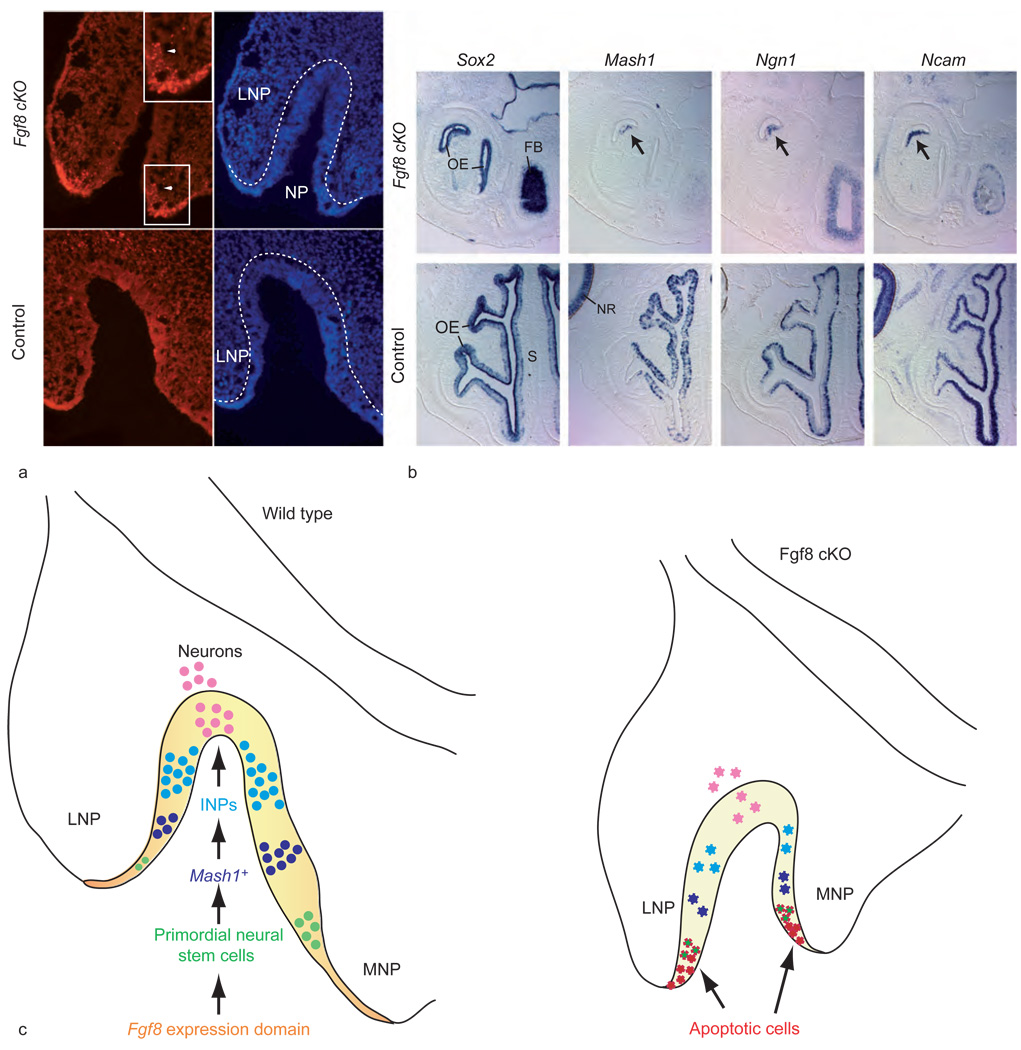
Fgf8 is required for cell survival in the neurogenic domain. (a) Terminal dUTP nick end labeling (TUNEL) of olfactory epithelium of mutant (cKO; conditional knockout) and control littermates at day 10.5 of gestation shows a high number of apoptotic cells in mutants in ectoderm and olfactory epithelium (white arrowhead; magnified in inset) of invaginating nasal pit (NP). Hoescht panel (blue, right) shows extent of invaginating NP. Broken white line indicates the boundary of the neuroepithelium lining the NP and lateral nasal process (LNP). (b) In situ hybridization on horizontal sections of mutant and control littermates (day 14.5 of gestation) shows the near absence of OE in the mutants and the lack of neuronal markers in the remaining OE (arrow) (FB, forebrain; S, nasal septum; NR, neural retina). (c) Role of Fgf8 in olfactory neurogenesis. Schematic of primary neurogenesis at day 10.5 of gestation in wild-type OE and Fgf8 mutant OE, illustrating the relative positions (MNP, medial nasal process) of the Fgf8 expression domain and different neuronal cell types: Fgf8 expression domain, orange; Sox2-expressing neuroepithelium, yellow; Sox2- and Fgf8-expressing primordial stem cells, green; Mash1-expressing progenitors, blue; immediate neuronal precursors (INPs),turquoise; Ncam-expressing olfactory receptor neurons, pink. In the mutant invaginating pit, cells undergoing apoptosis due to Fgf8 inactivation are shown in red and apoptotic primordial neural stem cells are green with red jagged borders. Diminished populations of other neuronal cell types are shown in their corresponding colors, but with jagged edges. Adapted from Kawauchi S, Shou J, Santos R, et al. (2005) Fgf8 expression defines a morphogenetic center required for olfactory neurogenesis and nasal cavity development in the mouse. Development 132(23): 5211–5223.
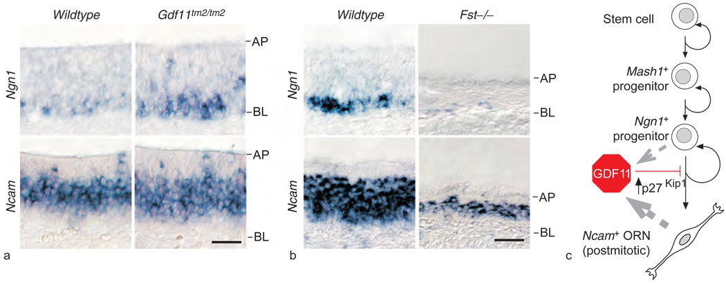
Disruption of neurogenesis in mice with loss or gain of function of Gdf11. (a) In situ hybridization on horizontal sections (AP, apical layer; BL, basal layer) of olfactory epithelium of Gdf11tm2/tm2 mice reveals an increase in Ngn1-expressing cells and a corresponding increase in Ncam-expressing cells: the Ncam-expressing cell layer is thicker by 20% (9 µm), about the diameter of one olfactory receptor neuron (ORN), in Gdf11tm2/tm2 olfactory epithelium, compared to in wild type. (b) Mice lacking a functional follistatin (Fst) gene show decreased olfactory epithelium neurogenesis. In situ hybridization for Ngn1 and Ncam shows large decreases in expression of both markers as well as aberrantly thin olfactory epithelium in the Fst mutant. (c) Schematic model of growth and differentiation factor 11 (GDF11) action in ORN neurogenesis. GDF11 is produced by both Ngn1+ progenitors and Ncam+ ORNs (gray broken-line arrows). GDF11 reversibly arrests Ngn1+ progenitors through induction of the cycle-dependent kinase inhibitor p27Kip1, thus preventing ORN generation. Reproduced from Wu HH, Ivkovic S, Murray RC, et al. (2003) Autoregulation of neurogenesis by GDF11. Neuron 37: 197–207, with permission from Elsevier.
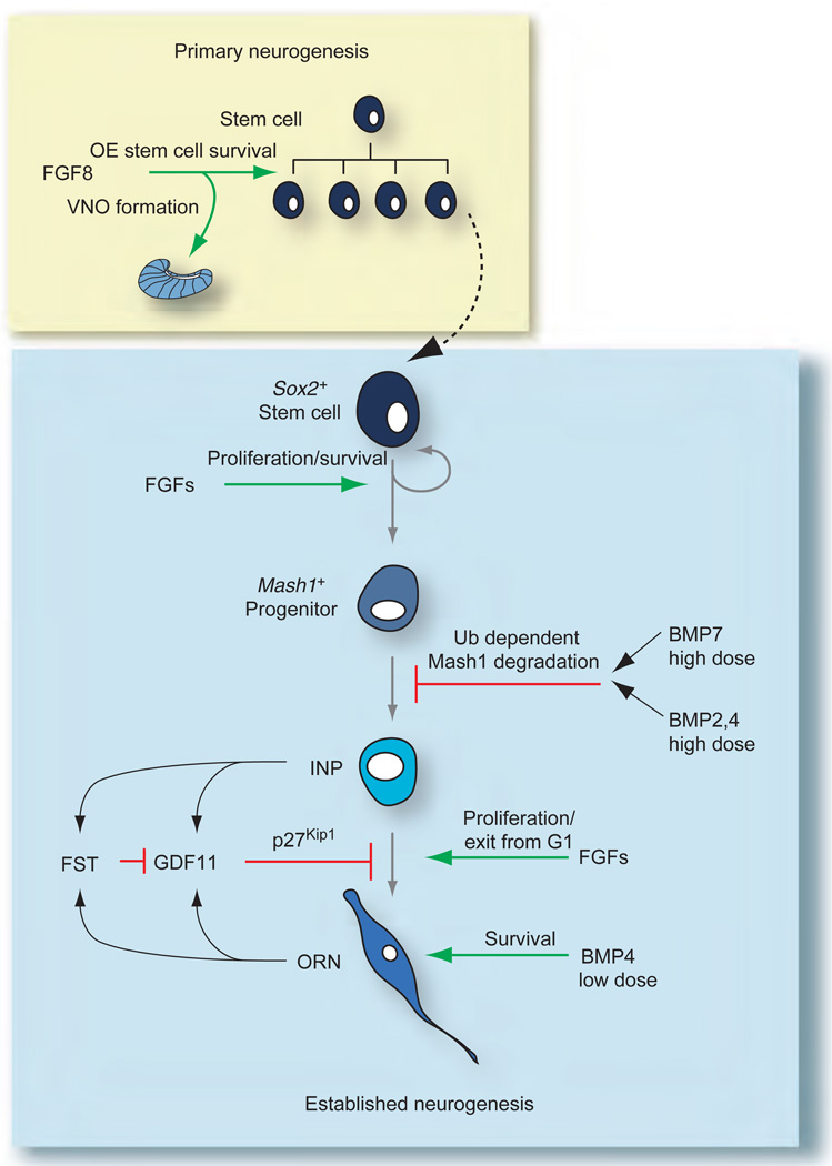
Summary of molecular signals involved in both primary and established olfactory epithelium (OE) neurogenesis. During primary neurogenesis (yellow box), fibroblast growth factor 8 (FGF8) and other factors are involved in producing the stem cell pool and initially establishing the different progenitors in the olfactory receptor neuron lineage. FGF8 is also necessary for initial vomeronasal organ (VNO) formation. After day 12.5 of gestation, once the lineage is established (blue box), FGFs and transforming growth factor-βs (BMP, bone morphogenetic protein; FST, follistatin; GDF, growth and differentiation factor) converge on different cell types in the OE lineage to achieve and maintain proper neuron number. Green arrows indicate stimulatory interactions; red bars indicate inhibitory interactions. Adapted from Kawauchi S, Beites CL, Crocker CE, et al. (2004) Molecular signals regulating proliferation of stem and progenitor cells in mouse olfactory epithelium. Developmental Neuroscience 26: 166–180, with permission. Copyright 2004 by S. Karger AG, Basel, Switzerland.
References
-
- Beites CL, Kawauchi S, Crocker CE, et al. Identification and molecular regulation of neural stem cells in the olfactory epithelium. Experimental Cell Research. 2005;306:309–316. - PubMed
-
- Bhattacharyya S, Bronner-Fraser M. Hierarchy of regulatory events in sensory placode development. Current Opinion in Genetics & Development. 2004;14:520–526. - PubMed
-
- Buck L, Axel R. A novel multigene family may encode odorant receptors: A molecular basis for odor recognition. Cell. 1991;65:175–187. - PubMed
-
- Caggiano M, Kauer JS, Hunter DD. Globose basal cells are neuronal progenitors in the olfactory epithelium: A lineage analysis using a replication-incompetent retrovirus. Neuron. 1994;13:339–352. - PubMed
-
- Cau E, Gradwohl G, Fode C, et al. Mash1 activates a cascade of bHLH regulators in olfactory neuron progenitors. Development. 1997;124:1611–1621. - PubMed
