. 2014 Mar 15;7(4):1337-47.
eCollection 2014.

Increasing the cytotoxicity of doxorubicin in breast cancer MCF-7 cells with multidrug resistance using a mesoporous silica nanoparticle drug delivery system

Affiliations

Increasing the cytotoxicity of doxorubicin in breast cancer MCF-7 cells with multidrug resistance using a mesoporous silica nanoparticle drug delivery system

Xin Wang et al. Int J Clin Exp Pathol. .

Abstract

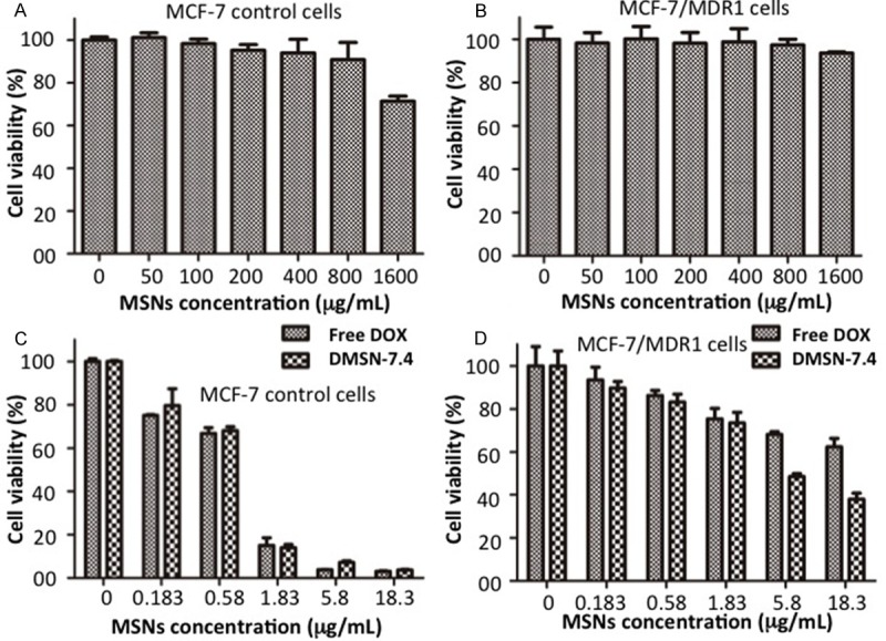
Resistance to cytotoxic chemotherapy is the main cause of therapeutic failure and death in women with breast cancer. Overexpression of various members of the superfamily of adenosine triphosphate binding cassette (ABC)-transporters has been shown to be associated with multidrug resistance (MDR) phenotype in breast cancer cells. MDR1 protein promotes the intracellular efflux of drugs. A novel approach to address cancer drug resistance is to take advantage of the ability of nanocarriers to sidestep drug resistance mechanisms by endosomal delivery of chemotherapeutic agents. Doxorubicin (DOX) is an anthracycline antibiotic commonly used in breast cancer chemotherapy and a substrate for ABC-mediated drug efflux. In the present study, we developed breast cancer MCF-7 cells with overexpression of MDR1 and designed mesoporous silica nanoparticles (MSNs) which were used as a drug delivery system. We tested the efficacy of DOX in the breast cancer cell line MCF-7/MDR1 and in a MCF-7/MDR1 xenograft nude mouse model using the MSNs drug delivery system. Our data show that drug resistance in the human breast cancer cell line MCF-7/MDR1 can be overcome by treatment with DOX encapsulated within mesoporous silica nanoparticles.

Keywords: Breast cancer; MDR; MSNs; drug delivery system.


Figures

Figure 1
Figure 1
High expression of MDR1 in MCF-7/MDR1 breast cancer cells. (A) MDR1 mRNA was expressed in MCF-7/MDR1 cells, but not in MCF-7 control cells. The house keeping gene GAPDH was used as an internal control of gene expression. (B) MDR1 protein was detected in MCF-7/MDR1 cells, but not in MCF-7 control cells. The house keeping gene-tubulin was used as an internal control. (C and D) MDR1 protein was not positively stained in MCF-7 control cells (C), but was positively stained in the cellular membrane and cytoplasm of MCF-7/MDR1 cells by immunohistochemical staining (D). (E and F) MDR1 protein was positively stained in the cellular membrane and cytoplasm in MCF-7/MDR1 cells using fluorescent immunohistochemical staining.
Figure 2
Figure 2
A. Transmission electron microscope image of MSNs exhibiting rod-shaped particles with a mean particle diameter of approximately 110 nm, uniform pore size of 2.7 nm and aspect ratio (AR) of 2.1-2.5. B. X-ray diffraction patterns of all the samples. The positions of the peaks represent the ordered hexagonal pore arrangements. C. The supernatant solutions of DMSN-5.4, DMSN-6.4, and DMSN-7.4 showed less color, respectively, indicating increased entrapment of DOX. D. The drug entrapment efficiency of DMSN-5.4, DMSN-6.4, and DMSN-7.4 increased by 25.2%, 63.8%, and 94.0%. There was a significant difference in entrapment of DOX with different solvent pH values (P<0.001).
Figure 3
Figure 3
Drug release efficacy was investigated in a physiological pH condition (PBS, pH 7.4) and acidic environment (pH 6.5 and 5.6). At pH 7.4, DMSNs have a low release rate of DOX, while at pH 5.6 and 6.5 the DOX release rate is high. The cumulative drug release rate for DMSN-7.4 reached 46.1% and 39.7% at pH 5.6 and 6.5, respectively.
Figure 4
Figure 4
A. Cellular internalization of DOX and DMSN-7.4 in MCF-7 and MCF-7/MDR1 cells was observed by fluorescence microscopy. B and C. The quantitative evaluation of free DOX and DMSN-7.4 was performed by photography under a fluorescence microscope (Olympus I×71, Japan) at ×400 magnification and measured with Image Pro Express software (Media Cybernetics, USA). D. TUNEL assay showing apoptosis by free DOX and DMSN-7.4 in MCF-7 cells and MCF-7/MDR cells after 24 h incubation.
Figure 5
Figure 5
Cytotoxicity of the MSNs was measured by MTT in both MCF-7 and MCF-7/MDR1 cells at different concentrations. A. MSNs exhibited limited toxicity with 71.4% cell viability of MCF-7 cells at 1600 μg/mL and negligible cytotoxicity at 800 μg/mL or lower. B. MSNs exhibited no obvious cytotoxicity in MCF-7/MDR1 cells at the concentrations of 50 to 1600 μg/mL even after culturing for 48 h. C. No significant difference in cell viability of MCF-7 control cells after exposure to different concentrations of free DOX and DMSN-7.4. D. There is a significant difference in the viability of MCF-7/MDR1 cells at the concentrations of 5.8 and 18.3 (mg/mL) (P<0.001).
Figure 6
Figure 6
The MSNs-DOX drug delivery system increases therapeutic efficacy in vivo. A. The free DOX group showed as small DOX signal similar to the saline group in the tumors, and the DMSN-7.4 group accumulated more DOX in the tumors. B. The free DOX group showed no significant tumor shrinkage. The DMSN-7.4 group had a significant therapeutic effect as indicated by the tumor weights (*P<0.05, **P<0.01).

References

    1. Ferlay J, Shin HR, Bray F, Forman D, Mathers C, Parkin DM. Estimates of worldwide burden of cancer in 2008: GLOBOCAN 2008. Int J Cancer. 2010;127:2893–2917. - PubMed
    1. Yang X, Uziely B, Groshen S, Lukas J, Israel V, Russell C, Dunnington G, Formenti S, Muggia F, Press MF. MDR1 gene expression in primary and advanced breast cancer. Lab Invest. 1999;79:271–280. - PubMed
    1. Zyad A, Benard J, Tursz T, Clarke R, Chouaib S. Resistance to TNF-alpha and adriamycin in the human breast cancer MCF-7 cell line: relationship to MDR1, MnSOD, and TNF gene expression. Cancer Res. 1994;54:825–831. - PubMed
    1. Liu F, Liu S, He S, Xie Z, Zu X, Jiang Y. Survivin transcription is associated with P-glycoprotein/ MDR1 overexpression in the multidrug resistance of MCF-7 breast cancer cells. Oncol Rep. 2010;23:1469–1475. - PubMed
    1. Xu D, Lu Q, Hu X. Down-regulation of P-glycoprotein expression in MDR breast cancer cell MCF-7/ADR by honokiol. Cancer Lett. 2006;243:274–280. - PubMed

Publication types

MeSH terms

Substances

LinkOut - more resources