

Small cell type neuroendocrine carcinoma colliding with squamous cell carcinoma at esophagus
- PMID: 24817981
- PMCID: PMC4014265
Small cell type neuroendocrine carcinoma colliding with squamous cell carcinoma at esophagus
Abstract
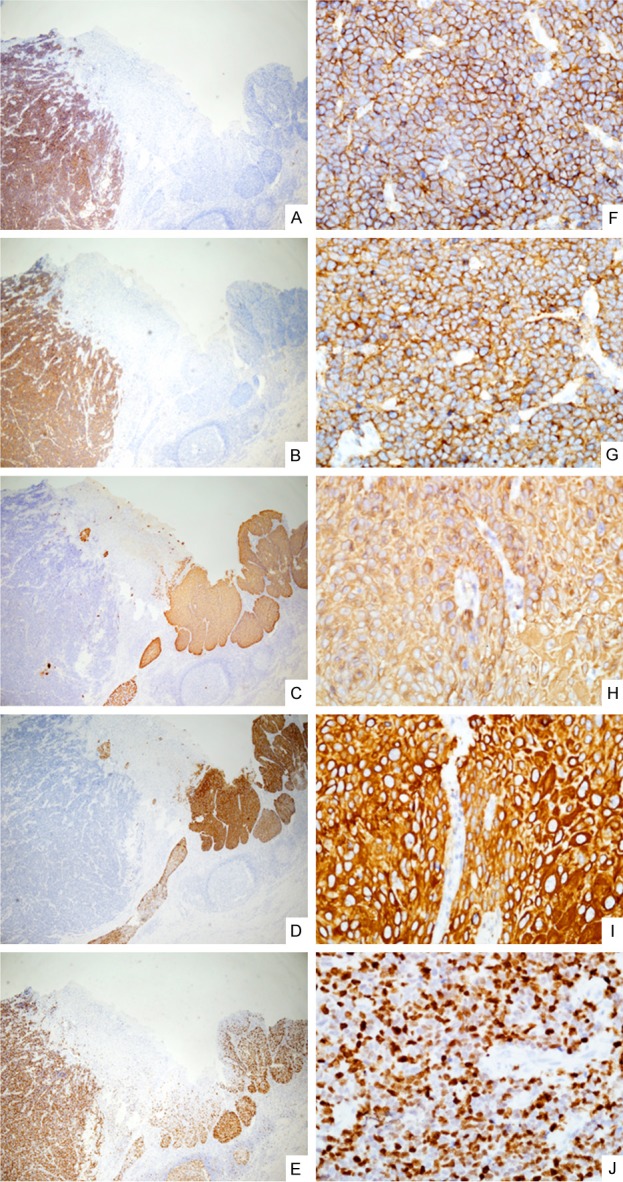
Collision tumor is an extremely rare tumor which defined as the concrescence of two distinct primaries neoplasms. We report here a case of collision tumor at lower third esophagus composed of small cell type neuroendocrine carcinoma (NEC), which is an very rare, highly aggressive and poorly prognostic carcinoma and squamous cell carcinoma (SqCC). In our case, pathologically, the small cell carcinoma display the characteristic of small, round, ovoid or spindle-shaped tumor cells with scant cytoplasm, which colliding with a moderately differentiated squamous cell carcinoma. Immunohistochemical staining demonstrated positive activities for CD56, synaptophysin, 34βE12, CK 5/6, ki-67 (70%-80%), but negative for CD99, chromogranin A, and TTF-1. Accurate diagnosis was made base on these findings.
Keywords: Collision tumor; esophagus; neuroendocrine carcinoma; small cell type; squamous cell carcinoma.
Figures

References
-
- Wittekind C, Yamasaki S. eds. Digestive system tumours. In: Sobin LH, Gospodarowicz MK, Wittekind C, editors. TNM Classification of Malignant Tumours. 7th Edition. Hoboken, PA: Wiley-Blackwell; 2009. pp. 63–135.
-
- Klöppel G. Classification and pathology of gastroenteropancreatic neuroendocrine neoplasms. Endocr Relat Cancer. 2011;18:S1–16. - PubMed
-
- Nakata S, Nagata Y, Sugaya M, Yasuda M, Yamashita T, Takenoyama M, Hanagiri T, Morita M, Hamada T, Sugio K, Yasumoto K. Primary pulmonary collision cancer consisting of large cell carcinoma and adenocarcinoma. Ann Thorac Surg. 2005;80:340–342. - PubMed
-
- Susnik B, Jordi Rowe J, Redlich PN, Chitambar C, Chang CC, Kampalath B. A unique collision tumor in breast: invasive ductal carcinoma and mucosa-associated lymphoid tissue lymphoma. Arch Pathol Lab Med. 2004;128:99–101. - PubMed
-
- Okumura K, Kato K, Furuhashi K, Suzuki K, Murase T. A collision cancer between urothelial carcinoma and malignant lymphoma of the urinary bladder: a case report. Hinyokika Kiyo. 2007;53:649–651. - PubMed
Publication types
MeSH terms
Substances
LinkOut - more resources
Full Text Sources
Medical
Research Materials