Review
. 2014 May 1:11:10.
doi: 10.1186/2045-8118-11-10. eCollection 2014.

A new look at cerebrospinal fluid circulation

Affiliations
Review

A new look at cerebrospinal fluid circulation

Thomas Brinker et al. Fluids Barriers CNS. .

Abstract

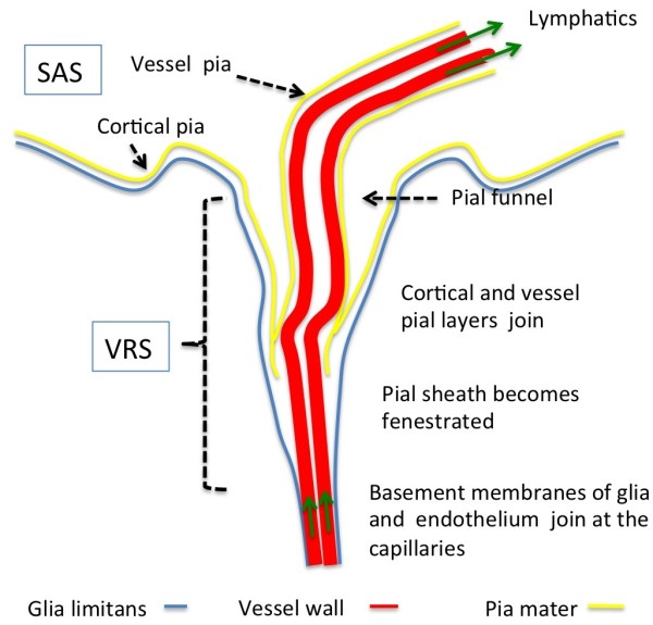
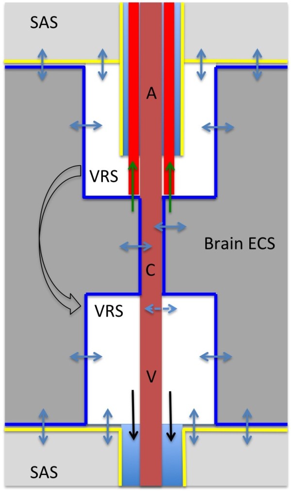
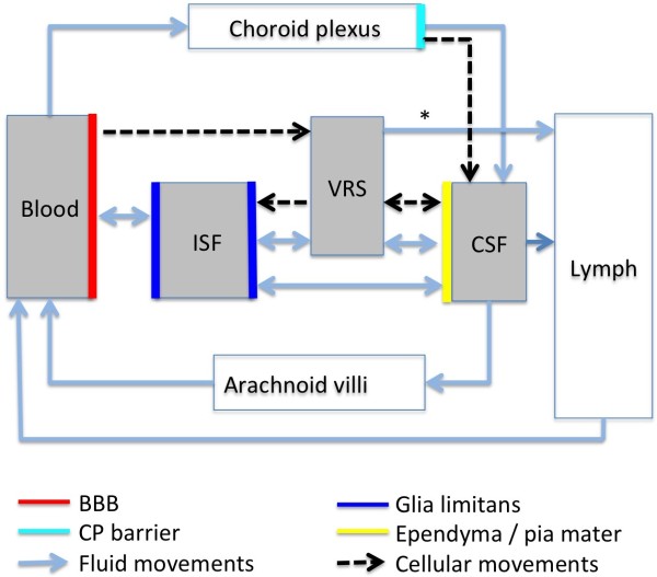
According to the traditional understanding of cerebrospinal fluid (CSF) physiology, the majority of CSF is produced by the choroid plexus, circulates through the ventricles, the cisterns, and the subarachnoid space to be absorbed into the blood by the arachnoid villi. This review surveys key developments leading to the traditional concept. Challenging this concept are novel insights utilizing molecular and cellular biology as well as neuroimaging, which indicate that CSF physiology may be much more complex than previously believed. The CSF circulation comprises not only a directed flow of CSF, but in addition a pulsatile to and fro movement throughout the entire brain with local fluid exchange between blood, interstitial fluid, and CSF. Astrocytes, aquaporins, and other membrane transporters are key elements in brain water and CSF homeostasis. A continuous bidirectional fluid exchange at the blood brain barrier produces flow rates, which exceed the choroidal CSF production rate by far. The CSF circulation around blood vessels penetrating from the subarachnoid space into the Virchow Robin spaces provides both a drainage pathway for the clearance of waste molecules from the brain and a site for the interaction of the systemic immune system with that of the brain. Important physiological functions, for example the regeneration of the brain during sleep, may depend on CSF circulation.

Keywords: Aquaporin; Astrocyte; Blood brain barrier; Cerebrospinal fluid circulation; Virchow Robin space.


Figures

Figure 1
Figure 1
Morphology of Virchow Robin and perivascular spaces. Delineated by basal membranes of glia, pia and endothelium, the Virchow Robin space (VRS) depicts the space surrounding vessels penetrating into the parenchyma. The VRS is obliterated at the capillaries where the basement membranes of glia and endothelium join. The complex pial architecture may be understood as an invagination of both cortical and vessel pia into the VRS. The pial funnel is not a regular finding. The pial sheath around arteries extends into the VRS, but becomes more fenestrated and eventually disappears at the precapillary section of the vessel. Unlike arteries (as shown in this figure), veins do not possess a pial sheath inside the VRS. ISF may drain by way of an intramural pathway along the basement membranes of capillaries and arterioles into the lymphatics at the base of the skull (green arrows). It should be noted that the figure does not depict the recently suggested periarterial flow from the SAS into the parenchyma and an outward flow into the cervical lymphatics along the veins (for discussion see text "Current research"). Also, it is still a matter of debate whether the Virchow Robin space, extending between the outer basement membrane of the vessel and the glia, represents a fluid-filled open space (see text). VRS: Virchow Robin space, SAS: subarachnoid space.
Figure 2
Figure 2
Diagram representing fluid movements at the Virchow Robin space. The complex anatomical structure of the Virchow Robin space (VRS) allows a bidirectional fluid exchange between the VRS and both the brain extracellular space (ECS) and the subarachnoid CSF space (blue arrows). Glial (blue lines) and pial (yellow lines) cell membranes enclose the VRS and control fluid exchange. Note, that it is a matter of debate whether the VRS represents an open fluid fill space (see text for discussion). Both experimental and clinical evidence indicate the existence of a pathway along the basement membranes of capillaries, arterioles, and arteries for the drainage of ISF and solutes into the lymphatic system (red lines and green arrows). It is unclear, whether the subpial perivascular spaces around arteries and veins (light blue) serve as additional drainage pathways. Also, the proposed glymphatic pathway connecting the arterial and venous VRS with the venous perivascular space (black arrows) is still a matter of debate. A: artery, V: vein, C: capillary, VRS: Virchow Robin space, SAS: subarachnoid space.
Figure 3
Figure 3
Diagram of the CSF "Circulation". This diagram summarizes fluid and cellular movements across the different barriers of the brain compartments (blood, interstitial fluid, Virchow Robin space, cerebrospinal fluid space comprising the cerebral ventricles, basal cisterns and cortical subarachnoid space). Aquaporins and other transporters control the fluid exchange at the glial, endothelial, and choroid plexus barrier. At the glial, endothelial, and pial barrier bi-directional flow may generate either a net in- or outflux, providing fluid exchange rates, which surpass the net CSF production rate by far. The choroid plexus is the only direct connection between the blood and the CSF compartment. Major portions of brain water are drained into the cervical lymphatics from the VRS (including its capillary section) via intramural arterial pathways (asterisks) and from the CSF space (via perineural subarachnoid space of cranial nerves). The capillary and venular endothelium may contribute to brain water absorption. Blood borne inflammatory cells may enter the brain via VRS venules or via CP. Fluid movements at the barriers are driven by osmotic and hydrostatic gradients or by active transporter processes. Fluid movements into and out of the VRS depend on respiratory and cardiac pressure pulsations.

References

    1. Milhorat TH. The third circulation revisited. J Neurosurg. 1975;42:628–645. doi: 10.3171/jns.1975.42.6.0628. - DOI - PubMed
    1. McComb JG. Recent research into the nature of cerebrospinal fluid formation and absorption. J Neurosurg. 1983;59:369–383. doi: 10.3171/jns.1983.59.3.0369. - DOI - PubMed
    1. Davson H. Formation and drainage of the cerebrospinal fluid. Sci Basis Med Annu Rev. 1966. pp. 238–259. - PubMed
    1. Johanson CE, Duncan JA, Klinge PM, Brinker T, Stopa EG, Silverberg GD. Multiplicity of cerebrospinal fluid functions: new challenges in health and disease. Cerebrospinal Fluid Res. 2008;5:10. doi: 10.1186/1743-8454-5-10. - DOI - PMC - PubMed
    1. Pardridge WM. Drug transport in brain via the cerebrospinal fluid. Fluids Barriers CNS. 2011;8:7. doi: 10.1186/2045-8118-8-7. - DOI - PMC - PubMed