. 2014 May 13:5:3812.
doi: 10.1038/ncomms4812.

Chd5 orchestrates chromatin remodelling during sperm development

Affiliations

Chd5 orchestrates chromatin remodelling during sperm development

Wangzhi Li et al. Nat Commun. .

Abstract

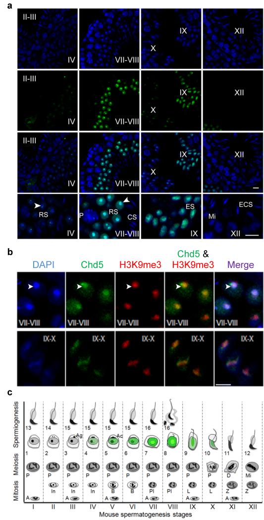
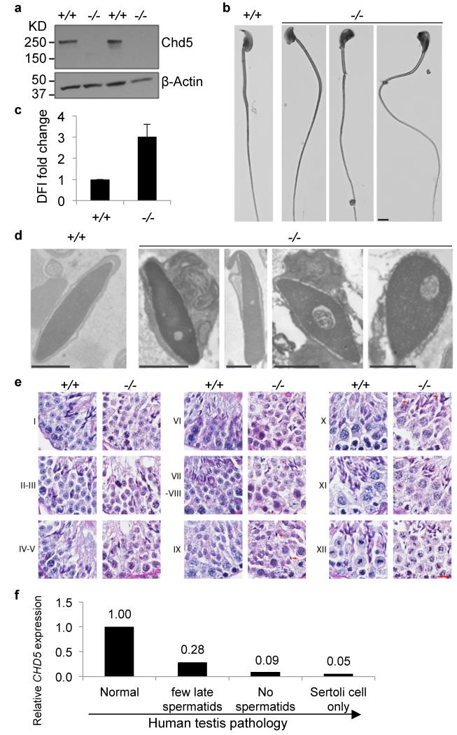
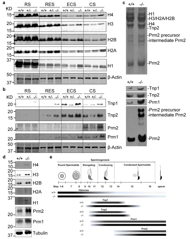
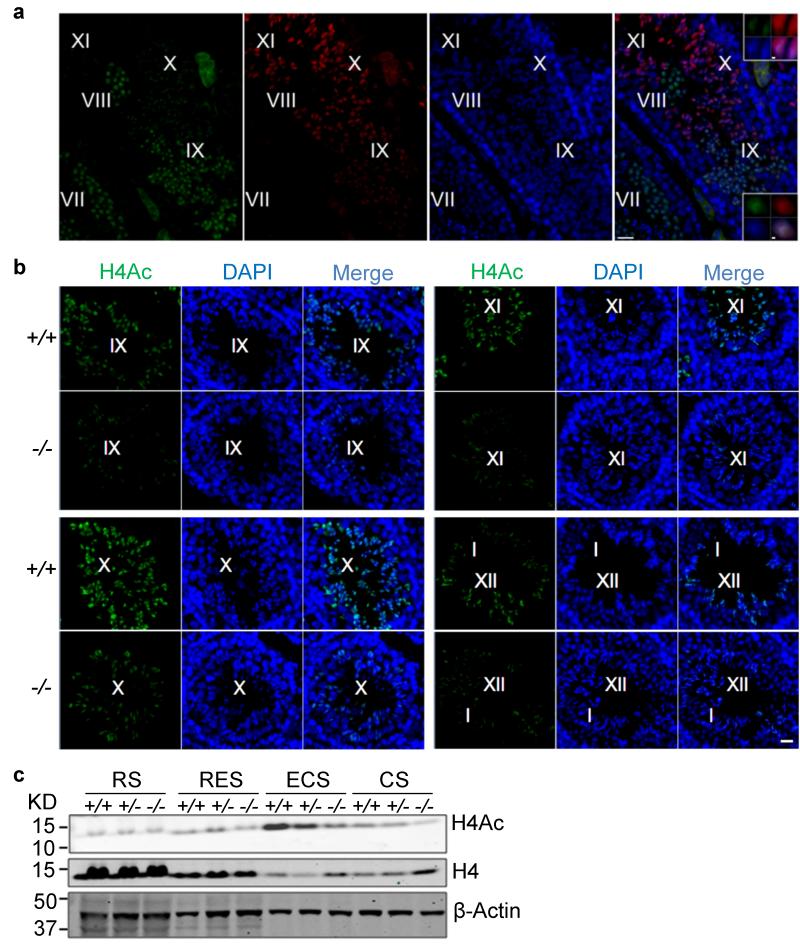
One of the most remarkable chromatin remodelling processes occurs during spermiogenesis, the post-meiotic phase of sperm development during which histones are replaced with sperm-specific protamines to repackage the genome into the highly compact chromatin structure of mature sperm. Here we identify Chromodomain helicase DNA binding protein 5 (Chd5) as a master regulator of the histone-to-protamine chromatin remodelling process. Chd5 deficiency leads to defective sperm chromatin compaction and male infertility in mice, mirroring the observation of low CHD5 expression in testes of infertile men. Chd5 orchestrates a cascade of molecular events required for histone removal and replacement, including histone 4 (H4) hyperacetylation, histone variant expression, nucleosome eviction and DNA damage repair. Chd5 deficiency also perturbs expression of transition proteins (Tnp1/Tnp2) and protamines (Prm1/2). These findings define Chd5 as a multi-faceted mediator of histone-to-protamine replacement and depict the cascade of molecular events underlying this process of extensive chromatin remodelling.


Figures

Figure 1
Figure 1. Chd5 is expressed in step 4 to 10 spermatids and is enriched in heterochromatin during spermiogenesis
Roman numerals indicate the spermatogenic stages of the tubules in wild type testes sections. (a) Blue, DAPI; Green, Chd5; RS, round spermatid; ES, elongating spermatid; ECS, elongating and condensing spermatid; CS, condensed spermatid; P, pachytene spermatocyte; Mi, meiotic division. Arrow heads mark the chromocenter. Scale bar, 10 μm. (b) Chd5 is enriched in DAPI-intense heterochromatic regions, and co-localizes with heterochromatin marker H3K9me3. Top panel, step 7-8 round spermatids; bottom panel, step 9-10 elongating spermatids. Arrow heads mark the chromocenter. Scale bar, 5 μm. (c) Schematic of Chd5 expression during spermatogenesis. Spermatogenesis is divided into twelve stages (stage I-XII) in mouse and each stage has a distinct cellular composition. Spermiogenesis, the maturation process of haploid spermatids, is divided into 16 steps (step 1-16). Green marks Chd5 protein expression. Chd5 is specifically expressed in spermatids from step 4 to 10, with peak expression in step 7-8 round spermatids, where Chd5 is enriched in the heterochromatic chromocenter. A focus of intense Chd5 protein expression is located adjacent to the junction of the chromocenter and the post-meiotic sex chromosomes in step 4-8 spermatids. Spermatogonia (A, In, B); Spermatocyte (Pl, preleptotene; L, leptotene; Z, zygotene; P, pachytene; D, diakinesis; Mi, meiotic division); Ag, acrosomic granule; Ac, acrosomic cap. The diagram is drawn based on the illustration of Dr. Rex Hess et al. in Chapter 1, Molecular Mechanisms in Spermatogenesis, Springer, 2008.
Figure 2
Figure 2. Chd5 deficiency leads to defective spermatogenesis and chromatin condensation
(a) Western blot analyses indicates that Chd5 protein is not detectable in Chd5Aam1−/− (−/−) testis. A validated antibody raised against a peptide for amino acids 1524-1705 of mouse Chd5 (which is not disrupted by gene targeting), was used for the Western blotting. β-Actin serves as a loading control. (b) Representative abnormal head morphology of Chd5Aam1−/− sperm. Scale bar, 20 μm. (c) SCSA revealed impaired chromatin integrity of Chd5Aam1−/− sperm. DFI, DNA Fragmentation Index (see Methods Summary). Data are presented as mean ± s.d. from four independent experiments. (d) Transmission electron microscopy analyses of sperm from Chd5Aam1+/+ and Chd5Aam1−/− caudal epididymi. Chromatin is homogenously condensed in Chd5Aam1+/+ sperm, but appears loose and uneven with fibrillar texture and contains abnormal vacuoles in Chd5Aam1−/− sperm nuclei. Scale bar, 1 μm. (e) Staged comparison of Periodic Acid-Schiff (PAS) stained Chd5Aam1+/+ and Chd5Aam1−/− testes. Roman numerals indicate the stages of the seminiferous tubules. A decrease in the number of elongated spermatids, especially at stage VII-VIII, is evident in Chd5Aam1−/− tubules. Arrows in stage IX and X mark abnormal retention of condensed spermatids. Scale bar, 1 μm. (f) Relative CHD5 expression in human testis with normal vs. clinically defined abnormal spermatogenesis. Data are derived from published microarray dataset (ArrayExpress: E-TABM-234) of 39 human testis biopsy samples from 29 men with highly defined testicular pathologies and 10 men with normal spermatogenesis. RNA was prepared from the testis biopsies and analyzed for gene expression using Affymetrix GeneChip. Data were analyzed through NextBio. Arrow indicates increased severity of spermatogenic defect.
Figure 3
Figure 3. Chd5 modulates histone removal and homeostasis of transition proteins and protamines
(a, b) Western blot analyses of protein lysates from purified spermatids at different spermiogenic stages. RS, round spermatids; RES, round and early elongating spermatids; ECS, elongating and condensing spermatids; CS, condensed spermatids. Increased retention of histones (H4, H3, H2B, H2A and H1) as well as elevated transition proteins (Tnp1, Tnp2) and protamines (Prm1, Prm2) are observed in elongating, condensing and condensed spermatids, but not in round spermatids of Chd5Aam1−/− (−/−) testis. β-Actin serves as a loading control. Levels of histones, transition proteins and protamines in Chd5Aam1+/- spermatids are in general similar to the levels in Chd5Aam1+/+ counterparts, with some variability among different Chd5Aam1+/- samples. The alterations in H3 in ECS, and alterations in Tnp2 and Prm2 in CS, are not consistently observed. (c) Top panel, Coomassie blue staining of basic protein extracts from Chd5Aam1+/+ and Chd5Aam1−/− sonication-resistant spermatids (SRS) separated using urea-acid gel electrophoresis. Equal amounts of proteins are loaded. Bottom panel, Western blot analyses of the same basic protein extracts show an increase in Tnp1, Tnp2, Prm1 and Prm2 in Chd5Aam1−/− SRS. The increase in the Prm2 precursor and intermediate Prm2 indicate deficient processing of the Prm2 precursor in Chd5Aam1−/− SRS. (d) Western blot analyses of protein lysates of sperm prepared from caudal epididymi of Chd5Aam1+/+ (+/+) and Chd5Aam1−/− (−/−) mice reveal increased retention of nucleosomal histones and elevated Prm1 and Prm2 in Chd5Aam1−/− sperm. H4, H2A and H1 are barely detectable in Chd5Aam1+/+ sperm, but are detected in Chd5Aam1−/− sperm. H3 and H2B are detectable in both Chd5Aam1+/+ and Chd5Aam1−/− sperm, but are higher in Chd5Aam1−/− sperm. Tubulin serves as a loading control. (e) Illustration of the dynamics of histones, transition proteins and protamines during spermiogenesis in Chd5Aam1+/+ (+/+) and Chd5Aam1−/− (−/−) testes. For each nuclear protein, the darker color of the bar in Chd5Aam1−/− testis indicates stronger expression of the protein at the indicated spermatogenic steps relative to Chd5Aam1+/+ counterparts.
Figure 4
Figure 4. Chd5 deficiency leads to compromised H4 acetylation during spermiogenesis
(a) Co-staining of Chd5 and H4 acetylation (H4Ac) in wild type testes. Roman numerals indicate spermatogenic stages of the marked tubular areas. Top and bottom inserts show higher magnification view of step 10 and step 9 spermatids, respectively. Both Chd5 and H4Ac are enriched in DAPI-intense regions within nuclei of step 9 and 10 spermatids in wild type testes. H4Ac is detected by a mouse monoclonal antibody against pan-H4K5/8/12/16Ac. Scale bars, 20 μm in main panels and 1 μm for inserts. (b) Immunofluorescence analyses of H4 acetylation in Chd5Aam1+/+ (+/+) and Chd5Aam1−/−(−/−) seminiferous tubules. Roman numerals indicate spermatogenic stages of the marked tubules. H4 hyperacetylation starts at step 9 spermatids of stage IX tubules, exhibits peak expression from step10 of stage X tubules to step 12 spermatids of stage XII tubules and decreases in step 13 spermatids in stage I tubules in Chd5Aam1+/+ (+/+) testis. H4 acetylation is weaker from step 9 to 13 spermatids in Chd5Aam1−/− (−/−) testis than in the Chd5Aam1+/+ counterparts. H4Ac was detected by a rabbit polyclonal antibody against pan-acetylation of H4. Scale bar, 20 μm. (c) Western blot analyses of purified spermatids at different spermiogenic stages. Histone H4 becomes transiently hyperacetylated in ECS of Chd5Aam1+/+ (+/+) testes, but not in ECS of Chd5Aam1−/− (−/−) testes. H4Ac was detected by a rabbit polyclonal antibody against pan-acetylation of H4. β-Actin serves as a loading control. RS, round spermatids; RES, round and early elongating spermatids; ECS, elongating and condensing spermatids; CS, condensed spermatids.
Figure 5
Figure 5. Chd5 deficiency leads to inefficient nucleosome eviction during spermiogenesis
Roman numerals indicate the spermatogenic stages of tubular areas. (a) Nucleosomes are detected in step 11 spermatids (indicated by arrow head) of stage XI seminiferous tubules, but are depleted afterwards and not detectable in step 12 spermatids (indicated by *) of stage XII seminiferous tubule in of wild type testes. Green, nucleosomes; Red, Lectin (visualizing acrosome for staging seminiferous tubules); Blue, DAPI; Mi, meiotic figure, a hallmark of stage XII tubules. (b) Immunostaining for nucleosomes show that nucleosomes are retained in as late as step 14 spermatids of stage II seminiferous tubules in Chd5Aam1−/− (−/−) testes, while absent in the Chd5Aam1+/+ (+/+) counterpart. Asterisks (*) mark nucleosome-positive condensing spermatids (step 14) in Chd5Aam1−/− tubules. Scale bar, 10 μm.
Figure 6
Figure 6. Increased DNA damage in Chd5Aam1−/− spermatids
(a) qRT-PCR analyses shows that Chd5 expression is induced approximately 5 fold when wild type mouse embryonic fibroblasts are treated with the DNA damaging agent adriamycin (0.4 μg/ml) for 24 hours. Results are normalized to Actb and the expression level at 0 hours is defined as 1. Data are presented as mean ± s.d. from three independent experiments. Adr, Adriamycin. (b) Increased TUNEL-positive condensing spermatids (step 13-15) in Chd5Aam1−/− testis. Data are presented as mean ± s.d. from three independent experiments. (c) Immunofluorescence analyses of γ-H2A.X in Chd5Aam1+/+ (+/+) and Chd5Aam1−/−(−/−) seminiferous tubules. Asterisks (*) mark γ-H2A.X-positive spermatids (step 13 in stage I and step 14 in stage II tubules) in Chd5Aam1−/− testis. Scale bar, 20 μm. (d) Western blot analyses of purified spermatids at different spermiogenic stages show a transient up-regulation of γ-H2A.X in wild type ECS, and increased accumulation of γ-H2A.X in Chd5Aam1−/− ECS and CS compared to wild type counterparts. β-Actin serves as a loading control. RS, round spermatids; RES, round and early elongating spermatids; ECS, elongating and condensing spermatids; CS, condensed spermatids.
Figure 7
Figure 7. Altered transcription of Prm1 and histone variants in Chd5-deficient spermatids
(a) qRT-PCR analyses of transition protein and protamine genes in Chd5Aam1+/+ and Chd5Aam1−/− round spermatids. Results are normalized to Actb expression. Prm1 expression is increased approximately 2.5 fold in Chd5Aam1−/− round spermatids. Data are presented as mean ± s.d. from four independent experiments. (b) Top panel, diagram of mouse Prm1 gene and location of primer sets used for ChIP-qPCR. Bottom panel, ChIP-qPCR analyses reveals enrichment of Chd5 at promoter region P (-77 bp to +135 bp) of Prm1 gene. The results are normalized to IgG control and are shown as fold of enrichment. Data are presented as mean ± s.d. from four to five independent experiments. (c) qRT-PCR analyses of rRNAs shows compromised expression of 28S and 45S ribosomal RNAs in Chd5Aam1−/− (−/−) round spermatids. Results are normalized to Actb expression. Data are presented as mean ± s.d. from four to five independent experiments. (d) qRT-PCR analyses of histone variants revealed an approximately 5 fold increase in expression of histone H2B variant Hist1h2bc and modest decreases in expression of histone variants Hist1h1e, Hist2h3c1 and H1t in Chd5Aam1−/− (−/−) round spermatids. Results are normalized to Actb expression. Data are presented as mean ± s.d. from three to five independent experiments.
Figure 8
Figure 8. RNA-Seq reveals global gene expression changes in Chd5Aam1−/− spermatids
(a) Heat map presentation of gene expression in Chd5Aam1+/+, Chd5Aam1+/- and Chd5Aam1−/− round spermatids revealed by RNA-Seq. Green to red (0 to 1) represents the gradient increase of expression levels. Sixty-five transcripts show upregulation, and 90 transcripts show downregulation, in Chd5Aam1−/− round spermatids. (b) qRT-PCR validation of RNA-Seq revealed gene expression changes in round spermatids. Results are normalized to Actb expression and data are presented as mean ± s.d. from four to five independent experiments. (c) Gene ontology analysis of the 155 transcripts that either show expression changes only in Chd5Aam1−/− spermatids but not in Chd5Aam1+/- spermatids, or that show gradual expression change from Chd5Aam1+/+ spermatids to Chd5Aam1+/- spermatids to Chd5Aam1−/− spermatids. Numbers next to bars indicate the number of genes classified to the corresponding GO term. P value is calculated using modified Fisher’s exact test by DAVID (v6.7). (d) qRT-PCR verification of compromised expression of genes implicated in histone acetylation, DNA damage response, RNA processing and nuclear integrity in Chd5Aam1−/− spermatids. Results are normalized to Actb expression and Data are presented as mean ± s.d. from four to five independent experiments.

Comment in

References

    1. Hess RA, Renato de Franca L. Spermatogenesis and cycle of the seminiferous epithelium. Advances in experimental medicine and biology. 2008;636:1–15. doi:10.1007/978-0-387-09597-4_1. - PubMed
    1. Tanaka H, Baba T. Gene expression in spermiogenesis. Cellular and molecular life sciences: CMLS. 2005;62:344–354. doi:10.1007/s00018-004-4394-y. - PMC - PubMed
    1. Oakberg EF. Duration of spermatogenesis in the mouse and timing of stages of the cycle of the seminiferous epithelium. The American journal of anatomy. 1956;99:507–516. doi:10.1002/aja.1000990307. - PubMed
    1. Oakberg EF. A description of spermiogenesis in the mouse and its use in analysis of the cycle of the seminiferous epithelium and germ cell renewal. The American journal of anatomy. 1956;99:391–413. doi:10.1002/aja.1000990303. - PubMed
    1. Ahmed EA, de Rooij DG. Staging of mouse seminiferous tubule cross-sections. Methods in molecular biology. 2009;558:263–277. doi:10.1007/978-1-60761-103-5_16. - PubMed

Publication types

Associated data