

Bone Marrow-Derived Stem Cell (BMDSC) transplantation improves fertility in a murine model of Asherman's syndrome
- PMID: 24819371
- PMCID: PMC4018329
- DOI: 10.1371/journal.pone.0096662
Bone Marrow-Derived Stem Cell (BMDSC) transplantation improves fertility in a murine model of Asherman's syndrome
Abstract
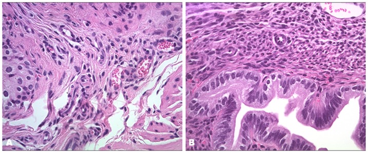
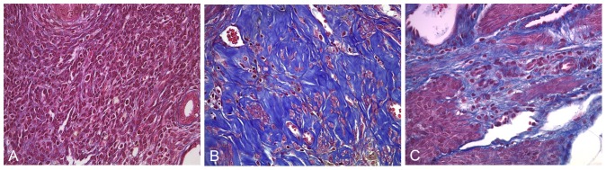
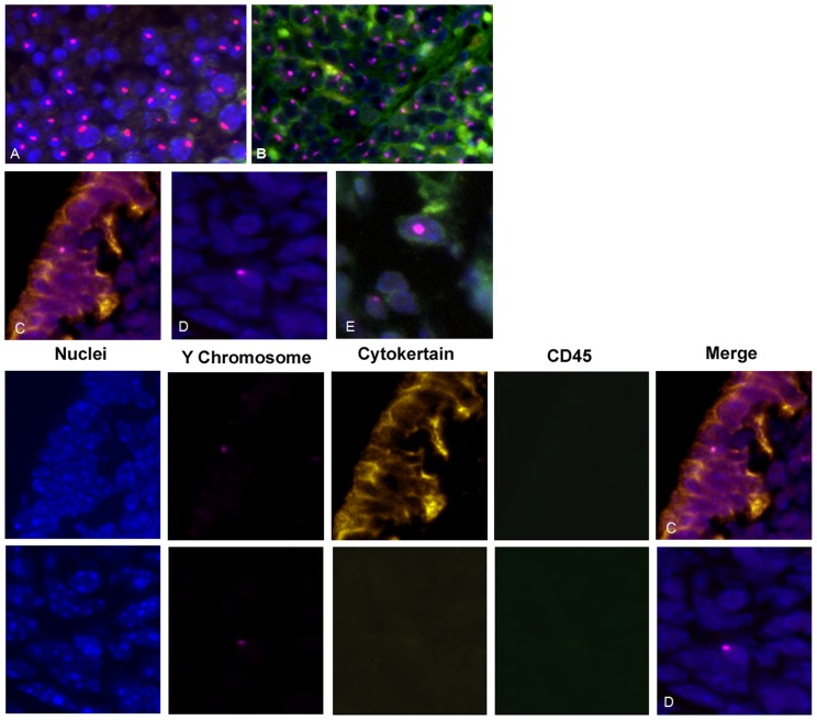
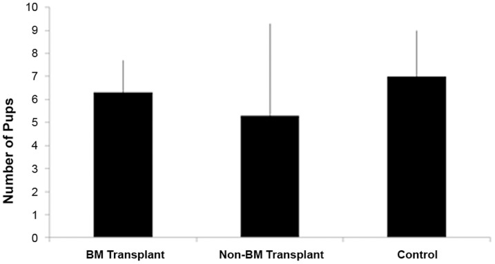
Asherman's Syndrome is characterized by intrauterine adhesions or fibrosis resulting as a consequence of damage to the basal layer of endometrium and is associated with infertility due to loss of normal endometrium. We have previously shown that bone marrow derived stem cells (BMDSCs) engraft the endometrium in mice and humans and Ischemia/reperfusion injury of uterus promoted BMDSCs migration to the endometrium; however, the role of BMDSCs in Asherman's syndrome has not been characterized. Here a murine model of Asherman's syndrome was created by traumatizing the uterus. We evaluate stem cell recruitment and pregnancy after BMDSCs transplantation in a model of Asherman's syndrome. In the Asheman's syndrome model, after BMDSC transplant, the Y chromosome bearing CD45-cells represented less than 0.1% of total endometrial cells. Twice the number of Y+CD45- cells was identified in the damaged uterus compared to the uninjured controls. There was no significant difference between the damaged and undamaged uterine horns in mice that received injury to a single horn. In the BMDSC transplant group, 9 of the 10 mice conceived, while only 3 of 10 in the non-transplanted group conceived (Chi-Square p = 0.0225); all mice in an uninjured control group conceived. The time to conception and mean litter size were not different between groups. Taken together, BMDSCs are recruited to endometrium in response to injury. Fertility improves after BMDSC transplant in Asherman's Syndrome mice, demonstrating a functional role for these cells in uterine repair. BMDSC transplantation is a potential novel treatment for Asherman's Syndrome and may also be useful to prevent Asherman's syndrome after uterine injury.
Conflict of interest statement
Figures
References
-
- Yu D, Wong Y, Cheong Y, Xia E, Li T (2008) Asherman syndrome—one century later. Fertil Steril 89: 759–779. - PubMed
-
- Tuuli MG, Shanks A, Bernhard L, Odibo AO, Macones GA, et al. (2012) Uterine synechiae and pregnancy complications. Obstet Gynecol 119: 810–814. - PubMed
-
- Buttram VC Jr, Turati G (1977) Uterine synechiae: variations in severity and some conditions which may be conducive to severe adhesions. Int J Fertil 22: 98–103. - PubMed
-
- Klein SM (1973) GarcíaCR (1973) Asherman's syndrome: a critique and current review. Fertil Steril 24: 722–735. - PubMed
-
- March CM (2011) Management of Asherman's syndrome. Reprod Biomed Online 23: 63–76. - PubMed
Publication types
MeSH terms
Grants and funding
LinkOut - more resources
Full Text Sources
Other Literature Sources
Medical
Research Materials
Miscellaneous
