Review
. 2014 Jul 11:16:53-76.
doi: 10.1146/annurev-bioeng-071813-104517. Epub 2014 Apr 16.

Computational modeling of cardiac valve function and intervention

Affiliations
Review

Computational modeling of cardiac valve function and intervention

Wei Sun et al. Annu Rev Biomed Eng. .

Abstract

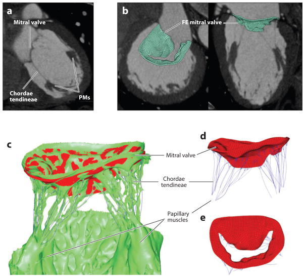
In the past two decades, major advances have been made in the clinical evaluation and treatment of valvular heart disease owing to the advent of noninvasive cardiac imaging modalities. In clinical practice, valvular disease evaluation is typically performed on two-dimensional (2D) images, even though most imaging modalities offer three-dimensional (3D) volumetric, time-resolved data. Such 3D data offer researchers the possibility to reconstruct the 3D geometry of heart valves at a patient-specific level. When these data are integrated with computational models, native heart valve biomechanical function can be investigated, and preoperative planning tools can be developed. In this review, we outline the advances in valve geometry reconstruction, tissue property modeling, and loading and boundary definitions for the purpose of realistic computational structural analysis of cardiac valve function and intervention.

Keywords: aortic valve; cardiac imaging; finite element analysis; heart valve; mitral valve.


Figures

Figure 1
Figure 1
Photographs of the structure of an excised human aortic root showing (a) the entire circumference of the annulus composed of fibrous and muscular regions and the coapting aortic leaflets—that is, the noncoronary (NCL), left coronary (LCL), and right coronary leaflets (RCL)—and (b) the same aortic root with the ascending aorta (AA), right (RCA) and left coronary arteries (LCA), noncoronary sinus of Valsalva (SOV), sinotubular junction (STJ), and trigone regions. (c) An illustration of the mitral valve anatomical structures, and (d) a photograph of an excised human mitral valve showing the mitral annulus, anterior (AML) and posterior mitral leaflets (PML), fibrous region, chordae tendineae (pink highlighted area), and papillary muscles (PMs).
Figure 2
Figure 2
(a) Drawing of the aortic valve showing a side view of one leaflet. (b) Schematic showing the side view of one leaflet in both the open and closed valve positions. Points A and C indicate the top of the commissures, point B (B′) indicates the middle point of the leaflet free edge in the open (closed) position, and point D indicates the middle point of the leaflet attachment line. Abbreviations: Db, diameter of the base; Dc, diameter of the commissures; H, valve height; Hs, height of the commissures; Lf, leaflet free-edge length; Lh, leaflet height; Xs, coaptation height in the center of the valve; α, angle of the closed valve; β, angle of the open valve; Ω, angle of the leaflet free edge in the open position. [Adapted with permission from Labrosse et al. (43).]
Figure 3
Figure 3
(top) Short-axis views of patient aortic valve computed tomography (CT) images at 20% (fully opened), 40%(half opened), and 80% (fully closed) of a cardiac cycle. (bottom) The corresponding reconstructed three-dimensional aortic root and leaflet finite element (FE) models showing fully opened, half-opened, and fully closed valve geometries (left to right).
Figure 4
Figure 4
Mean experimental biaxial response curves of (a) ovine, porcine, and human aortic leaflets in circumferential and radial directions and (b) porcine and ovine aortic sinuses and ascending aorta (AA) in circumferential and longitudinal directions.
Figure 5
Figure 5
A three-dimensional finite element (FE) mitral valve (MV) model. (a) The computed tomography (CT) long-axis two-chamber view of a closed MV showing a good visualization of chordae tendineae and papillary muscles (PMs). (b) The short-axis and long-axis views of the reconstructed FE MV model overlapped with the CT images. (c) The overlapping of geometries of the closed MV valve from the CT scans (green) and the simulated result (red) after applied pressure demonstrate a good match. Open MV valve geometry and the anatomical locations of chordae tendineae with chordal origins and papillary muscles are shown in (d) long-axis and (e) short-axis views.
Figure 6
Figure 6
A finite element model of a 94-year-old patient’s aorta, including the entire aortic root with coronary arteries, calcified leaflets, and a balloon-expandable transcatheter aortic valve (TAV) device. The simulation results predicted the aortic sinus rupture below the left coronary artery (LCA), which matched clinical observation.

References

    1. Thubrikar M. The Aortic Valve. Boca Raton, FL: CRC; 1990.
    1. Stella JA, Sacks MS. On the biaxial mechanical properties of the layers of the aortic valve leaflet. J Biomech Eng. 2007;129:757–66. - PubMed
    1. Jermihov PN, Jia L, Sacks MS, Gorman RC, Gorman JH, III, Chandran KB. Effect of geometry on the leaflet stresses in simulated models of congenital bicuspid aortic valves. Cardiovasc Eng Technol. 2011;2:48–56. - PMC - PubMed
    1. Conti C, Della Corte A, Votta E, Del Viscovo L, Bancone C, et al. Biomechanical implications of the congenital bicuspid aortic valve: a finite element study of aortic root function from in vivo data. J Thorac Cardiovasc Surg. 2010;140:890–96. - PubMed
    1. Katayama S, Umetani N, Sugiura S, Hisada T. The sinus of Valsalva relieves abnormal stress on aortic valve leaflets by facilitating smooth closure. J Thorac Cardiovasc Surg. 2008;136:1528–35. 1535.e1. - PubMed

LinkOut - more resources