

A spontaneous animal model of intestinal dysmotility evoked by inflammatory nitrergic dysfunction
- PMID: 24819503
- PMCID: PMC4018386
- DOI: 10.1371/journal.pone.0095879
A spontaneous animal model of intestinal dysmotility evoked by inflammatory nitrergic dysfunction
Abstract
Background and aims: Recent reports indicate the presence of low grade inflammation in functional gastrointestinal disorders (FGID), in these cases often called "post-inflammatory" FGIDs. However, suitable animal models to study these disorders are not available. The Biobreeding (BB) rat consists of a diabetes-resistant (BBDR) and a diabetes-prone (BBDP) strain. In the diabetes-prone strain, 40-60% of the animals develop diabetes and concomitant nitrergic dysfunction. Our aim was to investigate the occurrence of intestinal inflammation, nitrergic dysfunction and intestinal dysmotility in non-diabetic animals.
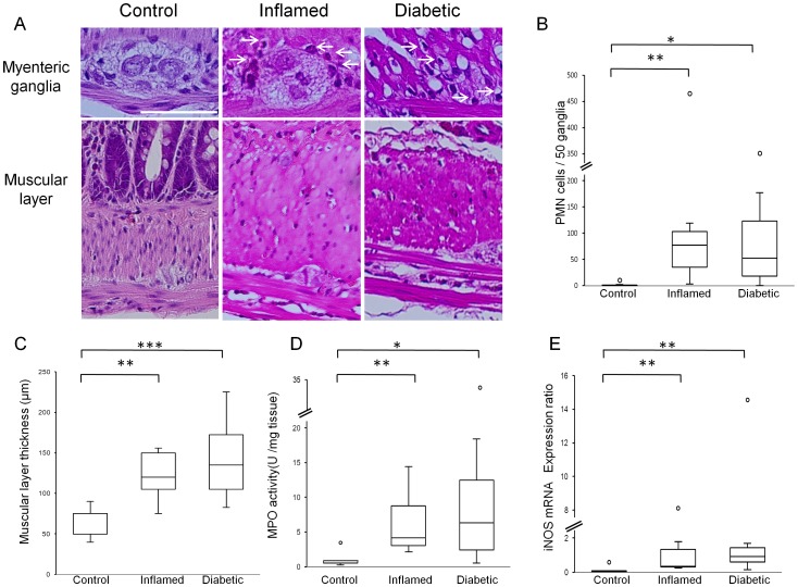
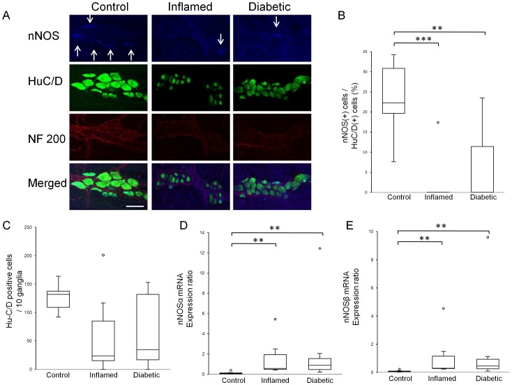
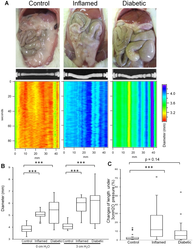
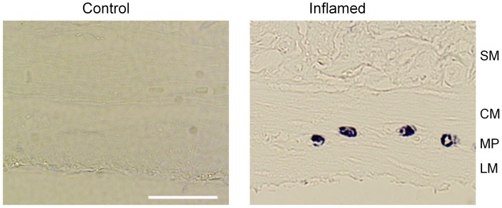
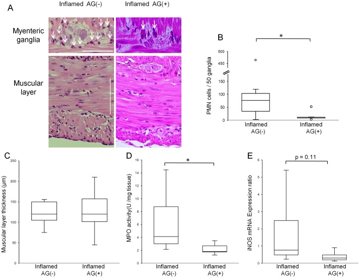
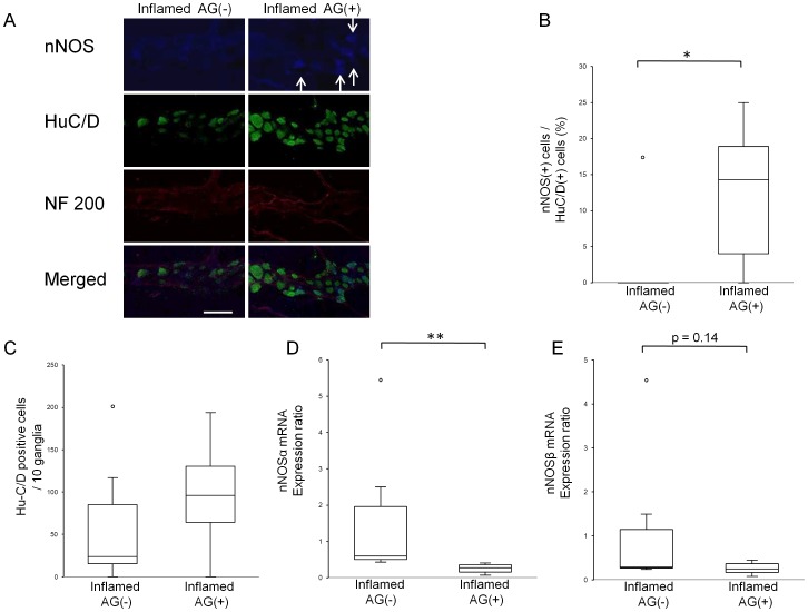
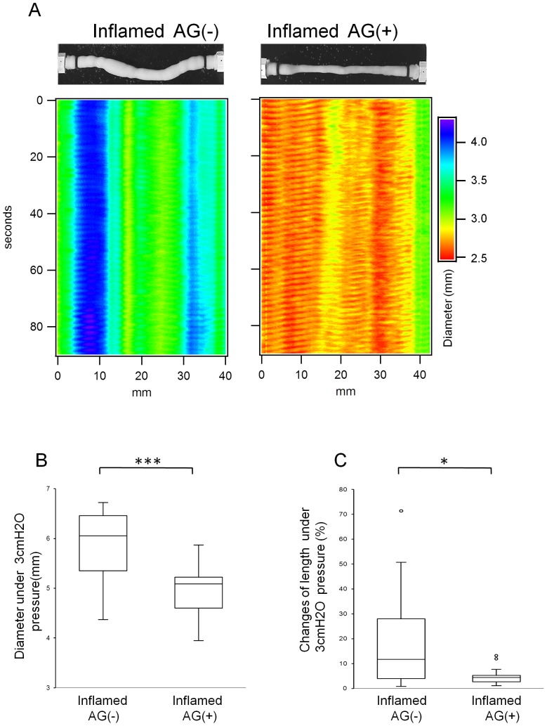
Methods: Jejunal inflammation (MPO assay, Hematoxylin&Eosin staining and inducible nitric oxide synthase (iNOS) mRNA expression), in vitro jejunal motility (video analysis) and myenteric neuronal numbers (immunohistochemistry) were assessed in control, normoglycaemic BBDP and diabetic BBDP rats. To study the impact of iNOS inhibition on these parameters, normoglycaemic BBDP rats were treated with aminoguanidine.
Results: Compared to control, significant polymorphonuclear (PMN) cell infiltration, enhanced MPO activity, increased iNOS mRNA expression and a decreased ratio of nNOS to Hu-C/D positive neurons were observed in both normoglycaemic and diabetic BBDP rats. Aminoguanidine treatment decreased PMN infiltration, iNOS mRNA expression and MPO activity. Moreover, it restored the ratio of nNOS to Hu-C/D positive nerves in the myenteric plexus and decreased the abnormal jejunal elongation and dilation observed in normoglycaemic BBDP rats.
Conclusions: Aminoguanidine treatment counteracts the inflammation-induced nitrergic dysfunction and prevents dysmotility, both of which are independent of hyperglycaemia in BB rats. Nitrergic dysfunction may contribute to the pathophysiology of "low-grade inflammatory" FGIDs. Normoglycaemic BBDP rats may be considered a suitable animal model to study the pathogenesis of FGIDs.
Conflict of interest statement
Figures
Similar articles
-
The normoglycaemic biobreeding rat: a spontaneous model for impaired gastric accommodation.Gut. 2016 Jan;65(1):73-81. doi: 10.1136/gutjnl-2014-308154. Epub 2014 Nov 19. Gut. 2016. PMID: 25410165
-
From intestinal permeability to dysmotility: the biobreeding rat as a model for functional gastrointestinal disorders.PLoS One. 2014 Oct 29;9(10):e111132. doi: 10.1371/journal.pone.0111132. eCollection 2014. PLoS One. 2014. PMID: 25354336 Free PMC article.
-
Nitric oxide regulates homeoprotein OTX1 and OTX2 expression in the rat myenteric plexus after intestinal ischemia-reperfusion injury.Am J Physiol Gastrointest Liver Physiol. 2017 Apr 1;312(4):G374-G389. doi: 10.1152/ajpgi.00386.2016. Epub 2017 Feb 2. Am J Physiol Gastrointest Liver Physiol. 2017. PMID: 28154013
-
Colonic hypersensitivity and low-grade inflammation in a spontaneous animal model for functional gastrointestinal disorders.Neurogastroenterol Motil. 2019 Jul;31(7):e13614. doi: 10.1111/nmo.13614. Epub 2019 May 8. Neurogastroenterol Motil. 2019. PMID: 31069897
-
Nitrergic Enteric Neurons in Health and Disease-Focus on Animal Models.Int J Mol Sci. 2019 Apr 24;20(8):2003. doi: 10.3390/ijms20082003. Int J Mol Sci. 2019. PMID: 31022832 Free PMC article. Review.
Cited by
-
Unravelling the Inflammatory Processes in the Early Stages of Diabetic Nephropathy and the Potential Effect of (Ss)-DS-ONJ.Int J Mol Sci. 2022 Jul 30;23(15):8450. doi: 10.3390/ijms23158450. Int J Mol Sci. 2022. PMID: 35955585 Free PMC article.
-
Persistent enteric neuroinflammation chronically impairs colonic motility in a pyridostigmine bromide-induced mouse model of Gulf War illness.Biol Open. 2025 Jun 15;14(6):bio061867. doi: 10.1242/bio.061867. Epub 2025 Jun 6. Biol Open. 2025. PMID: 40341359 Free PMC article.
-
Nitrergic and Substance P Immunoreactive Neurons in the Enteric Nervous System of the Bottlenose Dolphin (Tursiops truncatus) Intestine.Animals (Basel). 2021 Apr 8;11(4):1057. doi: 10.3390/ani11041057. Animals (Basel). 2021. PMID: 33918065 Free PMC article.
-
Serum Response Factor Is Essential for Prenatal Gastrointestinal Smooth Muscle Development and Maintenance of Differentiated Phenotype.J Neurogastroenterol Motil. 2015 Oct 1;21(4):589-602. doi: 10.5056/jnm15063. J Neurogastroenterol Motil. 2015. PMID: 26424044 Free PMC article.
-
Changes in Enteric Neurons of Small Intestine in a Rat Model of Irritable Bowel Syndrome with Diarrhea.J Neurogastroenterol Motil. 2016 Apr 30;22(2):310-20. doi: 10.5056/jnm15082. J Neurogastroenterol Motil. 2016. PMID: 26645247 Free PMC article.
References
-
- Drossman DA (2006) The functional gastrointestinal disorders and the Rome III process. Gastroenterology 130: 1377–90. - PubMed
-
- Kindt S, Tertychnyy A, de Hertogh G, Geboes K, Tack J (2009) Intestinal immune activation in presumed post-infectious functional dyspepsia. Neurogastroenterol Motil 21: 832–e56. - PubMed
-
- Dunlop SP, Hebden J, Campbell E, Naesdal J, Olbe L, et al. (2006) Abnormal intestinal permeability in subgroups of diarrhea-predominant irritable bowel syndromes. Am J Gastroenterol 101: 1288–94. - PubMed
-
- Tack J, Demedts I, Dehondt G Caenepeel P, Fischler B, et al. (2002) Clinical and pathophysiological characteristics of acute-onset functional dyspepsia. Gastroenterology 122: 1738–47. - PubMed
-
- Demedts I, Geboes K, Kindt S, Vanden Berghe P, Andrioli A, et al. (2006) Neural mechanisms of early postinflammatory dysmotility in rat small intestine. Neurogastroenterol Motil 18: 1102–11. - PubMed
Publication types
MeSH terms
Substances
LinkOut - more resources
Full Text Sources
Other Literature Sources
Research Materials
Miscellaneous
