. 2014 Jul;184(7):2111-22.
doi: 10.1016/j.ajpath.2014.04.002. Epub 2014 May 10.

Identification and characterization of a novel small-molecule inhibitor of β-catenin signaling

Affiliations

Identification and characterization of a novel small-molecule inhibitor of β-catenin signaling

Evan R Delgado et al. Am J Pathol. 2014 Jul.

Abstract

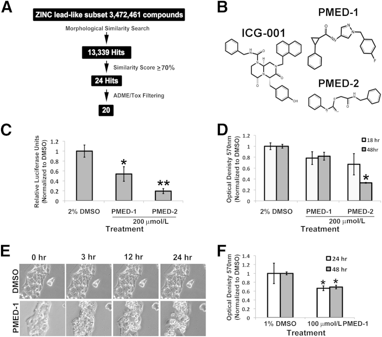
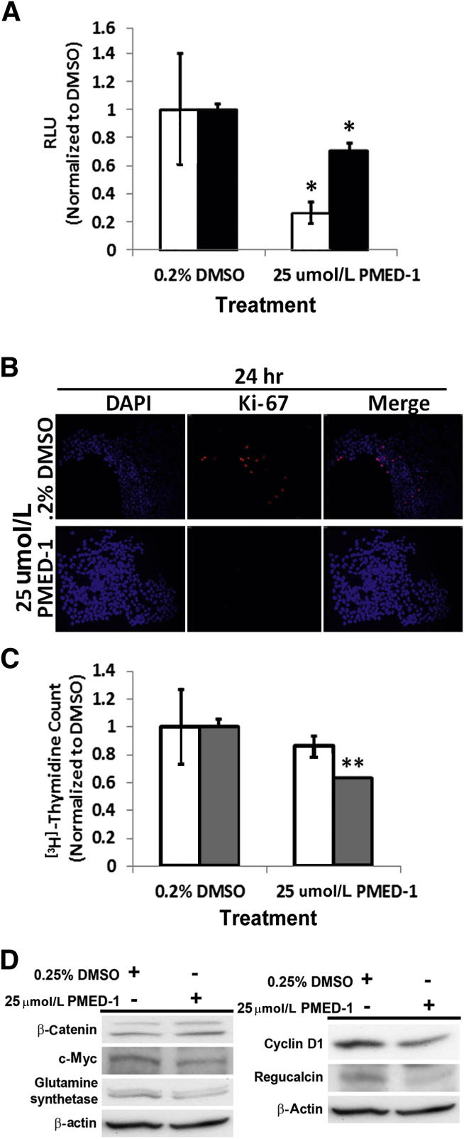
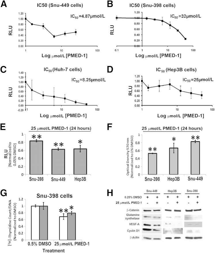
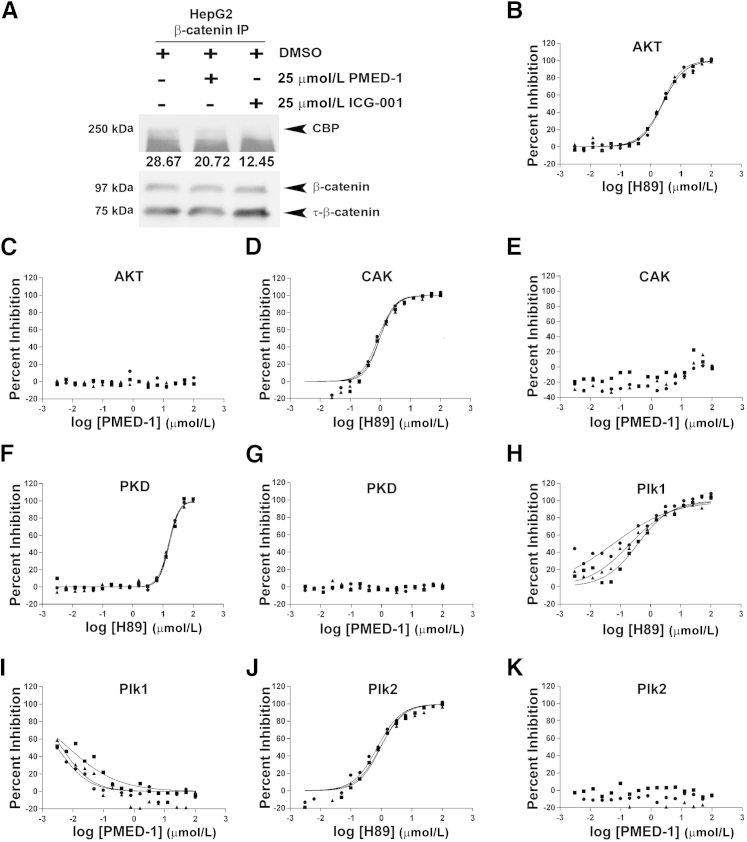
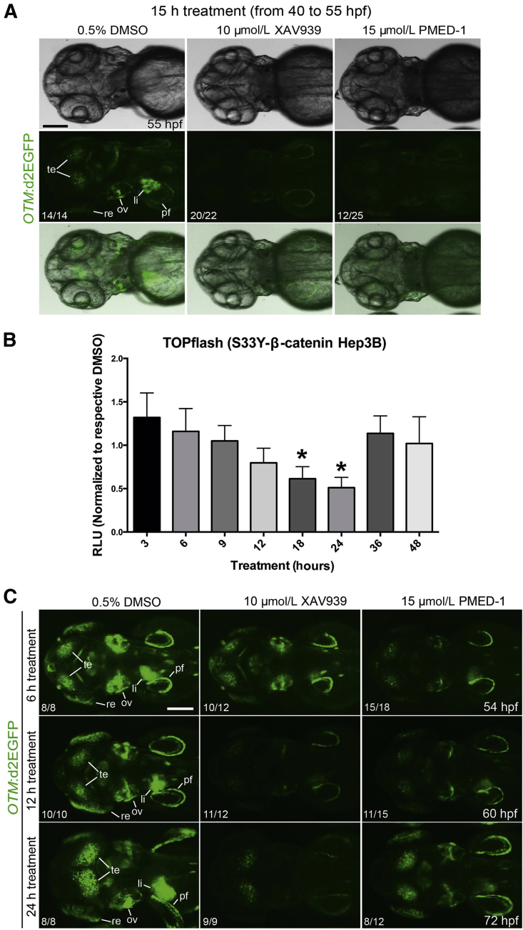
Hepatocellular carcinoma (HCC), the third most common cause of cancer-related deaths worldwide, lacks effective medical therapy. Large subsets of HCC demonstrate Wnt/β-catenin activation, making this an attractive therapeutic target. We report strategy and characterization of a novel small-molecule inhibitor, ICG-001, known to affect Wnt signaling by disrupting β-catenin-CREB binding protein interactions. We queried the ZINC online database for structural similarity to ICG-001 and identified PMED-1 as the lead compound, with ≥70% similarity to ICG-001. PMED-1 significantly reduced β-catenin activity in hepatoblastoma and several HCC cells, as determined by TOPflash reporter assay, with an IC50 ranging from 4.87 to 32 μmol/L. Although no toxicity was observed in primary human hepatocytes, PMED-1 inhibited Wnt target expression in HCC cells, including those with CTNNB1 mutations, and impaired cell proliferation and viability. PMED-1 treatment decreased β-catenin-CREB binding protein interactions without affecting total β-catenin levels or activity of other common kinases. PMED-1 treatment of Tg(OTM:d2EGFP) zebrafish expressing GFP under the β-catenin/Tcf reporter led to a notable decrease in β-catenin activity. The PMED effect on β-catenin signaling lasted from 12 to 24 hours in vitro and 6 to 15 hours in vivo. Thus, using a rapid and cost-effective computational methodology, we have identified a novel and specific small-molecule inhibitor of Wnt signaling that may have implications for HCC treatment.


Figures

Figure 1
Figure 1
Computational approach to identifying SMs that inhibit Wnt signaling to affect primary human hepatocytes and hepatic tumor cells. A: A search of the ZINC version 8.0 database lead-like subset identified 13,399 SMs structurally similar to ICG-001. A similarity score of ≥70% restricted the hits to 24 molecules, of which 20 passed in silico ADME/tox filtering. B: Chemical structure of ICG-001, PMED-1, and PMED-2. C: HepG2 cells treated with 200 μmol/L PMED-1 or PMED-2 for 24 hours exhibited a significant decrease in TOPflash luciferase reporter activity, compared with 2% DMSO. D: Primary hepatocytes treated with 2% DMSO or with 200 μmol/L PMED-1 or PMED-2 for 18 or 48 hours exhibited a significant decrease in viability by MTT assay only after PMED-2 treatment. E: HepG2 cells treated with 200 μmol/L PMED-1 for 24 hours monitored by live-cell imaging exhibit notable morphological changes reminiscent of cell death as early as 3 hours after treatment, compared with the 2% DMSO-treated controls. F: HepG2 cells treated with 100 μmol/L PMED-1 exhibited a significant decrease in cell viability as assessed by MTT assay, compared with 1% DMSO control. Data are expressed as means ± SD. P < 0.05, ∗∗P < 0.01. Original magnification, ×200 (E).
Figure 2
Figure 2
PMED-1 targets β-catenin signaling to affect cell proliferation in HepG2 cells. A: HepG2 cells treated with 25 μmol/L PMED-1 for 24 hours (white bars) exhibited a significant decrease in TOPflash reporter activity, but less so at 48 hours (black bars). B: Decrease in Ki-67+ cells (red) was observed after treatment of HepG2 cells with 25 μmol/L PMED-1 for 24 hours, compared with 0.2% DMSO. C: DNA synthesis in HepG2 cells as measured by [3H]thymidine incorporation exhibited a decrease in response to 24 (white bars) or 72 (gray bars) hours of treatment with 25 μmol/L of PMED-1, compared with 0.2% DMSO. D: Western blots using HepG2 cell lysates reveal decreased β-catenin targets after 25 μmol/L PMED-1 treatment. Data are expressed as means ± SD. P < 0.05, ∗∗P < 0.01. Original magnification, ×100 (B).
Figure 3
Figure 3
IC50 of PMED-1 in various HCC cells and the effect of PMED-1 on β-catenin signaling and biology. A–D: IC50 was determined by response of TOPflash reporter activity to PMED-1 treatment for 24 hours in Snu-449 (A), Snu-398 (B), Huh-7 (C), and Hep3B (D) cells. E: Relative decrease in TOPflash reporter activity after 25 μmol/L PMED-1 treatment of various HCC cells for 24 hours. F: The most robust decrease in viability as measured by MTT assay was observed in Snu-398 cells after 24 hours of 25 μmol/L PMED-1 treatment. G: DNA synthesis as measured by [3H]thymidine incorporation was significantly reduced in Snu-398 cells treated for 24 (white bars) or 72 (gray bars) hours with 25 μmol/L PMED-1, compared with DMSO. H: Western blots using whole-cell lysates from Snu-398, Snu-449, and Hep3B cells treated with 25 μmol/L PMED-1 reveal notable decreases in β-catenin downstream targets. Data are expressed as means ± SD. P < 0.05, ∗∗P < 0.01.
Figure 4
Figure 4
PMED-1 affects β-catenin–CBP interactions but does not affect activity of other common kinases. A: Western blot analysis of whole-cell lysates from untreated HepG2 cells or HepG2 cells treated for 24 hours with either 2% DMSO, 25 μmol/L PMED-1, or 25 μmol/L ICG-001 used for immunoprecipitation revealed significantly less CBP pull-down with anti–β-catenin antibody in cells treated with PMED-1 or ICG-001, compared with DMSO. Densitometry values (arbitrary units) for CBP are provided under the bands. B–K: Effect of H89 and PMED-1 on activity of kinases as assessed by IMAP-based FP assay (AKT and PKD) or TR-Fret assay (CAK, Plk1, and Plk2). Three replicates are shown. B: H89 decreases CAK activity in a dose-dependent manner. C: PMED-1 does not affect AKT activity. D: H89 decreases CAK activity. E: PMED-1 does not affect CAK activity. F: H89 decreases PKD activity at higher concentrations. G: PMED-1 does not affect PKD activity. H: H89 decreases Plk1 activity in a largely dose-dependent manner. I: PMED-1 had an inhibitory effect on Plk1 up to −1 μmol/L on the log scale (0.097 μmol/L); however, no decrease was evident at higher concentrations. J: H89 decreases Plk2 activity in a dose-dependent manner. K: PMED-1 does not affect Plk2 activity. IP, immunoprecipitation.
Figure 5
Figure 5
Efficacy and duration of the effect of PMED-1 effect on β-catenin activity in vivo and in vitro. A: GFP+ embryos obtained from outcrossing Tg(OTM:d2EGFP) zebrafish were treated with DMSO, XAV939, or PMED-1 for 15 hours (40 to 55 hpf), and GFP expression was assessed under a fluorescence microscope. Shown are bright-field images (top row), GFP fluorescence images (middle row), and merged images (bottom row). B: Stably transfected Hep3B cells with S33Y-β-catenin, treated with a single dose of PMED-1 at 25 μmol/L, exhibited a gradual decrease in TOPflash reporter activity; the decrease reached significance at 18 and 24 hours after treatment, with rebound at 36 and 48 hours. C: Epifluorescence images of GFP expression in Tg(OTM:d2EGFP) embryos. Embryos strongly GFP+ [obtained from incrossing Tg(OTM:d2EGFP) zebrafish] were treated with DMSO, XAV939, or PMED-1 from 48 hpf. Embryos were harvested 6, 12, or 24 hours later, and GFP expression was assessed under a fluorescence microscope. P < 0.05. The proportion of embryos (n/N) exhibiting the GFP expression is indicated at the lower left of each image. All images are dorsal view, with anterior to the left. Scale bars: 100 μm (A and C). li, liver; ov, otic vesicle; pf, pectoral fin; re, retina; te, tectum.

Similar articles

Cited by

References

    1. Ferlay J., Shin H.R., Bray F., Forman D., Mathers C., Parkin D.M. Estimates of worldwide burden of cancer in 2008: GLOBOCAN 2008. Int J Cancer. 2010;127:2893–2917. - PubMed
    1. Yang J.D., Roberts L.R. Hepatocellular carcinoma: a global view. Nat Rev Gastroenterol Hepatol. 2010;7:448–458. - PMC - PubMed
    1. Fortune B.E., Umman V., Gilliland T., Emre S. Liver transplantation for hepatocellular carcinoma: a surgical perspective. J Clin Gastroenterol. 2013;47(Suppl):S37–S42. - PubMed
    1. Imamura H., Matsuyama Y., Tanaka E., Ohkubo T., Hasegawa K., Miyagawa S., Sugawara Y., Minagawa M., Takayama T., Kawasaki S., Makuuchi M. Risk factors contributing to early and late phase intrahepatic recurrence of hepatocellular carcinoma after hepatectomy. J Hepatol. 2003;38:200–207. - PubMed
    1. Suzuki K., Kono N., Ono A., Osuga Y., Kiyokawa H., Mineo I., Matsuda Y., Miyoshi S., Kawata S., Minami Y., Moriwaki K., Tarui S. Transcatheter arterial chemo-embolization for humoral hypercalcemia of hepatocellular carcinoma. Gastroenterol Jpn. 1988;23:29–36. - PubMed

Publication types

MeSH terms