. 2014 May 12;9(5):e96778.
doi: 10.1371/journal.pone.0096778. eCollection 2014.

The use of Nanotrap particles technology in capturing HIV-1 virions and viral proteins from infected cells

Affiliations

The use of Nanotrap particles technology in capturing HIV-1 virions and viral proteins from infected cells

Elizabeth Jaworski et al. PLoS One. .

Abstract

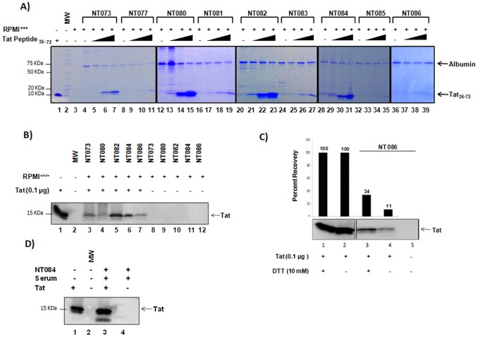
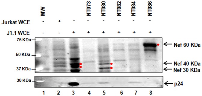
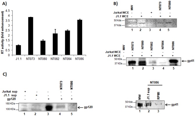
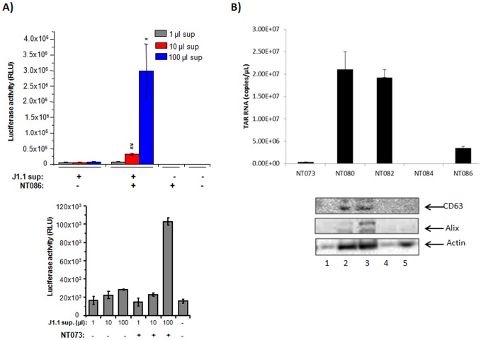
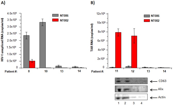
HIV-1 infection results in a chronic but incurable illness since long-term HAART can keep the virus to an undetectable level. However, discontinuation of therapy rapidly increases viral burden. Moreover, patients under HAART frequently develop various metabolic disorders and HIV-associated neuronal disease. Today, the main challenge of HIV-1 research is the elimination of the residual virus in infected individuals. The current HIV-1 diagnostics are largely comprised of serological and nucleic acid based technologies. Our goal is to integrate the nanotrap technology into a standard research tool that will allow sensitive detection of HIV-1 infection. This study demonstrates that majority of HIV-1 virions in culture supernatants and Tat/Nef proteins spiked in culture medium can be captured by nanotrap particles. To determine the binding affinities of different baits, we incubated target molecules with nanotrap particles at room temperature. After short sequestration, materials were either eluted or remained attached to nanotrap particles prior to analysis. The unique affinity baits of nanotrap particles preferentially bound HIV-1 materials while excluded albumin. A high level capture of Tat or Tat peptide by NT082 and NT084 particles was measured by western blot (WB). Intracellular Nef protein was captured by NT080, while membrane-associated Nef was captured by NT086 and also detected by WB. Selective capture of HIV-1 particles by NT073 and NT086 was measured by reverse transcriptase assay, while capture of infectious HIV-1 by these nanoparticles was demonstrated by functional transactivation in TZM-bl cells. We also demonstrated specific capture of HIV-1 particles and exosomes-containing TAR-RNA in patients' serum by NT086 and NT082 particles, respectively, using specific qRT-PCR. Collectively, our data indicate that certain types of nanotrap particles selectively capture specific HIV-1 molecules, and we propose to use this technology as a platform to enhance HIV-1 detection by concentrating viral proteins and infectious virions from infected samples.


Conflict of interest statement

Competing Interests: BL is employed by Ceres Nanosciences. LL and EP hold patents on the described nanoparticle enrichment technology invented at George Mason University (NIH grant #'s 5R33CA173359, 5R21A1099851, 5R21CA137706) and Licensed by Ceres Nanosciences. Patent details are: United States 7,935,518 B2 Utility (CIP) Smart Hydrogel Particles for Biomarker Harvesting, EP 1929299 National Stage Methods of Isolating Analytes from a Sample, JP 494753 National Stage Methods of Isolating Analytes from a Sample, United States 8,382,987 B2 Utility (CIP) Method for Harvesting Nanoparticles and Sequestering Biomarkers, US 8,497,137 B2 Utility (CIP) Smart Hydrogel Particles for Biomarker Harvesting, EP 2132256 National Stage Smart Hydrogel Particles for Biomarker Harvesting. Co-authors, Lance Liotta, Emanuel Petricoin and Fatah Kashanchi are members of the Scientific Advisory board at Ceres Nanosciences. LL, EP and BL are share holders at Ceres Nanosciences. The nanoparticles used in this study were research grade and provided by Ceres Nanosciences which are not commercially available products. This does not alter the authors' adherence to all of the PLOS ONE policies on sharing data and materials.

Figures

Figure 1
Figure 1. Capture of Tat protein by nanotrap particles.
(A) Tat36–72 peptide was spiked into RPMI-1640 containing FBS, antibiotics and glutamine (RPMI+++) at 0.1 µg (lanes 5, 9, 13, 17, 21, 25, 29, 33, 37), 1 µg (lanes 6, 10, 14, 18, 22, 26, 30, 34, 38), and 10 µg (lanes 7, 11, 15, 19, 23, 27, 31, 35, 39). A 30% slurry of nanotrap particles prepared in unsupplemented RPMI was incubated with RPMI containing Tat36–72 for 30 min at room temperature. Eluted materials (15 µl) were then coomassie stained for the presence of captured Tat peptide. (B) Full-length Tat protein spiked in RPMI-1640 (+++) (100 µl) was captured by a 30% slurry of each nanotrap particle in unsupplemented RPMI (lanes 3–7), separated on SDS-PAGE and detected by Western blot using anti-tat antibody. Nanotrap particles incubated with RPMI+++ without Tat were used as background control (lanes 8–12). (C) Full-length Tat was pre-treated with DTT at 37°C for 30 min (lanes 1 and 3) and incubated with NT086 (lane 3). Percent recovery Tat capture under reducing conditions was determined from raw densitometry counts of the western blot. (D) Tat protein (1 µg) was spiked into uninfected patient serum (100 µl), incubated with NT084 (lane 3), and subsequent Tat capture determined by western blot.
Figure 2
Figure 2. Capture of HIV-1 Nef and p24 capsid proteins by nanotrap particles.
Aliquots of infected J1.1 whole cell extract (WCE) (100 µg) in 100 µl of water was incubated with 75 µl of a 30% slurry of nanotrap particles in TNE buffer without detergent for 30 min (lanes 4–8). Nef binding capacity of each nanotrap particle was subsequently determined by western blot using α-Nef antibody (upper panel). The binding of p24 to nanotrap particles was also determined by western blot using α-p24 antibody (lower panel). Both Jurkat and J1.1 WCE were used as background controls (lanes 2, 3) without enrichment by nanotrap particles.
Figure 3
Figure 3. Capture of HIV-1 virions by nanotrap particles.
(A) A 30% slurry of each nanotrap particle in unsupplemented RPMI (30 µl) was incubated with J1.1 cell supernatants (200 µl) for 30 min at room temperature. Virus-bound nanoparticles were washed to remove unbound virus, and the nanoparticle pellets were suspended in 50 µl of Tris (250 nM) + 1% Triton X-100, and assayed for RT activity without elution. Fold enrichment of virus binding to nanotrap particles was analyzed over J1.1 supernatant alone without prior enrichment. (B) HIV-1 gp41 was detected in J1.1 WCE by WB without nanoparticles pretreatment (upper panel, lane 2). The uninfected Jurkat WCE was used as negative control (lane 3). Binding of HIV-1 by nanotrap particles through interaction with gp41 was detected in J1.1 WCE following treatments with nanoparticles NT073 and NT080 (lanes 4 and 5, respectively). The captures of gp41 from J1.1 WCE were also shown by nanoparticles NT073, NT080 and NT086 but not by NT084 (middle panel). Fresh antibody was used for this panel. (C) Interaction of nanotrap particles with HIV-1 envelope gp120 was detected by western blot using anti-HIV human serum. Briefly, purified gp120 glycoprotein (1 µg) or J1.1 culture supernatant was spiked into water and incubated with or without nanotrap particles slurry for 30 min at room temperature, washed and suspended in Laemmli buffer before resolved on SDS-PAGE gel and analyzed by western blot.
Figure 4
Figure 4. Capture of infectious HIV-1 virions by nanotrap particles.
(A) HIV-1 infected J1.1 supernatants (1, 10, 100 µl) diluted to 1 ml in complete media, were either untreated or added to 50 µl of 30% slurry of NT086 nanotrap particles. The particles were incubated for one hour with gentle rotation at room temperature. The unbound virus was removed by centrifugation for 10 min before incubating the nanotrap particles with TZM-bl cells at 37°C in microtiter plates. Virus samples were also incubated with TZM-bl cells without prior treatment with nanotrap particles. After 48 hr post incubation, cells were lysed and HIV-1 transactivation analyzed by luciferase assay. The 2 asterices (p<0.01) and 1 asterix (p<0.05) represent the level of statistical significance between virus captures from 1 and 10 µl, and 10 and 100 µl supernatants, respectively. The NT073 nanoparticles were similarly used to capture infectious virions from J1.1 supernatants and analyzed by TZM-bl system as above (B) Exosomes (15 µl) collected from J1.1 cell supernatant were spiked into PBS (85 µl) and then incubated with a 30% slurry of five different nanotrap particles (30 µl) for 30 min. Nanotrap particles were washed and subjected to Trizol buffer for total RNA extraction. Evaluation of TAR-RNA contents of exosomes captured by nanotrap particles was performed via qRT-PCR using specific TAR-RNA primers. The five different nanotrap particles were similarly incubated with infected J1.1-derived exosomes and washed as in panel B. The nanoparticles-bound exosomes were then suspended in Laemmli buffer, separated on SDS-PAGE gel and analyzed by western blot using antibody to CD63 and Alix (standard markers for exosomes).
Figure 5
Figure 5. Capture of HIV-1 virions and exosomes containing TAR-RNA in patients' serum by nanotrap particles.
Aliquots of serum samples from 4 HIV-1 infected HAART individuals (#8, #10, #11 and #12) and 2 uninfected donors (#13 and #14) were incubated separately with nanotrap particles (NT086 and NT082) for 30 min at room temperature. After removing unbound materials by washing, nanotrap particles were incubated with Trizol buffer for total RNA extraction. The levels of HIV-1 virions and exosomes containing TAR-RNA bound to each type of nanotrap particles were quantified by qRT-PCR using primers specific to HIV-1 unspliced RNA (A) and TAR-RNA (B). The similarly prepared nanotrap particles-bound serum-derived materials (from samples #11, #12, #13 and #14) were also suspended in Laemmli buffer, separated on SDS-PAGE gel, and analyzed by western blot using antibody to CD63 and Alix (standard markers for exosomes) (see lower panel).
Figure 6
Figure 6. HIV-1 and exosomes capturing capacity of nanoparticles.
(A) 100 µl aliquot of the dual-tropic HIV-1 89.6 containing 10 or 1 ng p24/ml was incubated with a pellet of 0.02, 0.2 or 2.0 mg of NT086 for 30 min at room temperature. The virus bound-nanoparticles were washed and RNA isolated. The RNA was converted to cDNA and PCR reaction mixtures were prepared using high-low concentrations of cDNA, Gag primers and the iTaq Universal SYBR Green Supermix. Serial dilutions of DNA from 8E5 cells were used as the standards. Quantitative real-time PCR reactions were carried out in triplicate. The asterix represents the statistical significance at the level of p<0.05. (B) The infected J1.1 cells were treated with or without anti-retrovirals (ART), and then sups were incubated with NT086 to capture virions. After washing, the total RNA was extracted from the nanoparticle captured materials and then subjected to qRT-PCR using specific primers to unspliced HIV-1 RNA. (C) The infectivity of the nanoparticles captured virions were also analyzed by incubating them with TZM-bl cells and measuring luciferase activity. The asterix represents the statistical significance at the level of p<0.05. (D) The NT080 nanoparticles capture of exosomal RNA from J1.1 supernatants with or without pretreatment of ART were similarly measured by qRT-PCR using specific primers to TAR RNA.
Figure 7
Figure 7. Schematic diagram of a nanotrap particle.
The outer porous shell, inner core and the affinity baits in the core are represented by yellow, blue and green, respectively. In some nanotrap particles vinyl sulfonic acid (VSA) is incorporated into the outer shell. The smaller viral proteins readily enter into the particle core through the pores of the shell and bind to the specific affinity baits. The larger viral particles do not completely enter into the core of the nanoparticle, and thus bind to the outer shell of the particles.

Similar articles

Cited by

References

    1. Fields BN KD, Howley PM, et al... (2013) Fields Virology 6th Ed - Human Immunodeficiency Virus: Philadelphia: Wolters Kluwer Health/Lippincott Williams & Wilkins.
    1. Varghese B, Maher JE, Peterman TA, Branson BM, Steketee RW (2002) Reducing the risk of sexual HIV transmission: quantifying the per-act risk for HIV on the basis of choice of partner, sex act, and condom use. Sex Transm Dis 29: 38–43. - PubMed
    1. Pilcher CD, Eron JJ Jr, Galvin S, Gay C, Cohen MS (2004) Acute HIV revisited: new opportunities for treatment and prevention. J Clin Invest 113: 937–945. - PMC - PubMed
    1. Donegan E, Stuart M, Niland JC, Sacks HS, Azen SP, et al. (1990) Infection with human immunodeficiency virus type 1 (HIV-1) among recipients of antibody-positive blood donations. Ann Intern Med 113: 733–739. - PubMed
    1. Ndirangu J, Viljoen J, Bland RM, Danaviah S, Thorne C, et al. (2012) Cell-free (RNA) and cell-associated (DNA) HIV-1 and postnatal transmission through breastfeeding. PLoS One 7: e51493. - PMC - PubMed

Publication types

Substances