

Cardioprotective trafficking of caveolin to mitochondria is Gi-protein dependent
- PMID: 24821070
- PMCID: PMC4219636
- DOI: 10.1097/ALN.0000000000000295
Cardioprotective trafficking of caveolin to mitochondria is Gi-protein dependent
Abstract
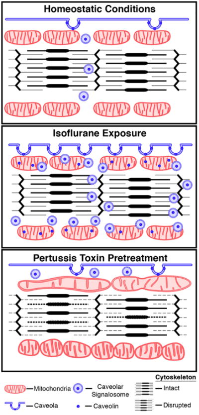
Background: Caveolae are a nexus for protective signaling. Trafficking of caveolin to mitochondria is essential for adaptation to cellular stress though the trafficking mechanisms remain unknown. The authors hypothesized that G protein-coupled receptor/inhibitory G protein (Gi) activation leads to caveolin trafficking to mitochondria.
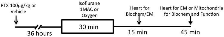
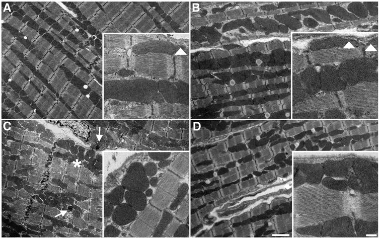
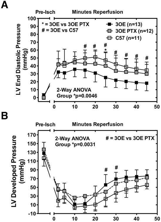
Methods: Mice were exposed to isoflurane or oxygen vehicle (30 min, ± 36 h pertussis toxin pretreatment, an irreversible Gi inhibitor). Caveolin trafficking, cardioprotective "survival kinase" signaling, mitochondrial function, and ultrastructure were assessed.
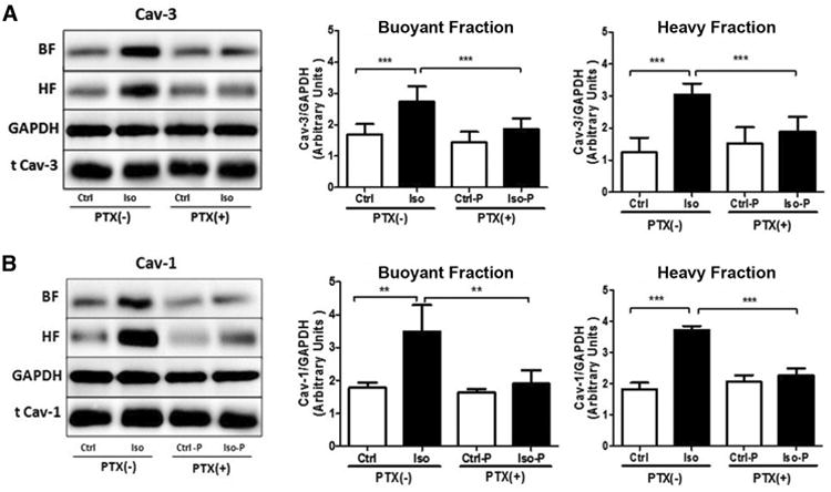
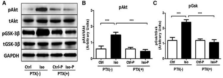
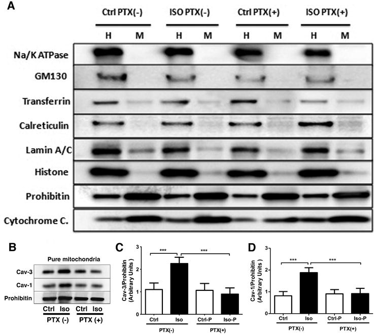
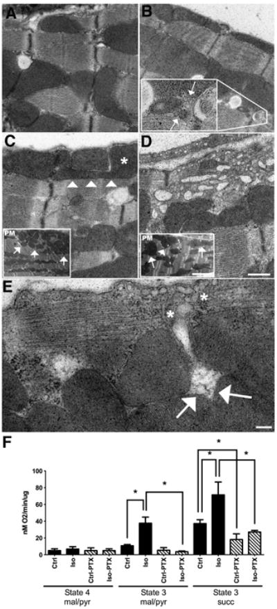
Results: Isoflurane increased cardiac caveolae (n = 8 per group; data presented as mean ± SD for Ctrl versus isoflurane; [caveolin-1: 1.78 ± 0.12 vs. 3.53 ± 0.77; P < 0.05]; [caveolin-3: 1.68 ± 0.29 vs. 2.67 ± 0.46; P < 0.05]) and mitochondrial caveolin levels (n = 16 per group; [caveolin-1: 0.87 ± 0.18 vs. 1.89 ± .19; P < 0.05]; [caveolin-3: 1.10 ± 0.29 vs. 2.26 ± 0.28; P < 0.05]), and caveolin-enriched mitochondria exhibited improved respiratory function (n = 4 per group; [state 3/complex I: 10.67 ± 1.54 vs. 37.6 ± 7.34; P < 0.05]; [state 3/complex II: 37.19 ± 4.61 vs. 71.48 ± 15.28; P < 0.05]). Isoflurane increased phosphorylation of survival kinases (n = 8 per group; [protein kinase B: 0.63 ± 0.20 vs. 1.47 ± 0.18; P < 0.05]; [glycogen synthase kinase 3β: 1.23 ± 0.20 vs. 2.35 ± 0.20; P < 0.05]). The beneficial effects were blocked by pertussis toxin.
Conclusions: Gi proteins are involved in trafficking caveolin to mitochondria to enhance stress resistance. Agents that target Gi activation and caveolin trafficking may be viable cardioprotective agents.
Figures
Similar articles
-
Role of G(i/o)-Src kinase-PI3K/Akt pathway and caveolin-1 in β₂-adrenoceptor coupling to endothelial NO synthase in mouse pulmonary artery.Cell Signal. 2011 Jul;23(7):1136-43. doi: 10.1016/j.cellsig.2011.02.008. Epub 2011 Mar 6. Cell Signal. 2011. PMID: 21385608
-
Cardiac-specific overexpression of caveolin-3 induces endogenous cardiac protection by mimicking ischemic preconditioning.Circulation. 2008 Nov 4;118(19):1979-88. doi: 10.1161/CIRCULATIONAHA.108.788331. Epub 2008 Oct 20. Circulation. 2008. PMID: 18936328 Free PMC article.
-
Mechanistic insights into δ-opioid-induced cardioprotection: Involvement of caveolin translocation to the mitochondria.Life Sci. 2020 Apr 15;247:116942. doi: 10.1016/j.lfs.2019.116942. Epub 2019 Nov 9. Life Sci. 2020. PMID: 31715185
-
The caveolin triad: caveolae biogenesis, cholesterol trafficking, and signal transduction.Cytokine Growth Factor Rev. 2001 Mar;12(1):41-51. doi: 10.1016/s1359-6101(00)00022-8. Cytokine Growth Factor Rev. 2001. PMID: 11312118 Review.
-
The Caveolin genes: from cell biology to medicine.Ann Med. 2004;36(8):584-95. doi: 10.1080/07853890410018899. Ann Med. 2004. PMID: 15768830 Review.
Cited by
-
Helium-Induced Changes in Circulating Caveolin in Mice Suggest a Novel Mechanism of Cardiac Protection.Int J Mol Sci. 2019 May 29;20(11):2640. doi: 10.3390/ijms20112640. Int J Mol Sci. 2019. PMID: 31146391 Free PMC article.
-
Shear stress augments mitochondrial ATP generation that triggers ATP release and Ca2+ signaling in vascular endothelial cells.Am J Physiol Heart Circ Physiol. 2018 Nov 1;315(5):H1477-H1485. doi: 10.1152/ajpheart.00204.2018. Epub 2018 Aug 24. Am J Physiol Heart Circ Physiol. 2018. PMID: 30141983 Free PMC article.
-
Caveolin-3 deficiency associated with the dystrophy P104L mutation impairs skeletal muscle mitochondrial form and function.J Cachexia Sarcopenia Muscle. 2020 Jun;11(3):838-858. doi: 10.1002/jcsm.12541. Epub 2020 Feb 23. J Cachexia Sarcopenia Muscle. 2020. PMID: 32090499 Free PMC article.
-
Altered Penile Caveolin Expression in Diabetes: Potential Role in Erectile Dysfunction.J Sex Med. 2017 Oct;14(10):1177-1186. doi: 10.1016/j.jsxm.2017.08.006. J Sex Med. 2017. PMID: 28923309 Free PMC article.
-
Caveolins in cardioprotection - translatability and mechanisms.Br J Pharmacol. 2015 Apr;172(8):2114-25. doi: 10.1111/bph.13009. Epub 2015 Jan 13. Br J Pharmacol. 2015. PMID: 25377989 Free PMC article. Review.
References
-
- Hausenloy DJ, Yellon DM. Survival kinases in ischemic preconditioning and postconditioning. Cardiovasc Res. 2006;70:240–53. - PubMed
-
- Sanada S, Komuro I, Kitakaze M. Pathophysiology of myocardial reperfusion injury: Preconditioning, postconditioning, and translational aspects of protective measures. Am J Physiol Heart Circ Physiol. 2011;301:H1723–41. - PubMed
-
- Palade G. Fine structure of blood capillaries. J Appl Physiol. 1953;24:1424.
-
- Pike LJ. Lipid rafts: Bringing order to chaos. J Lipid Res. 2003;44:655–67. - PubMed
Publication types
MeSH terms
Substances
Grants and funding
- R01 NS073653/NS/NINDS NIH HHS/United States
- HL091071/HL/NHLBI NIH HHS/United States
- HL107200/HL/NHLBI NIH HHS/United States
- I01 BX001963/BX/BLRD VA/United States
- R01 HL115933/HL/NHLBI NIH HHS/United States
- I01 BX001225/BX/BLRD VA/United States
- R01 HL091071/HL/NHLBI NIH HHS/United States
- HL115933/HL/NHLBI NIH HHS/United States
- HL066941/HL/NHLBI NIH HHS/United States
- NS073653/NS/NINDS NIH HHS/United States
- R01 HL107200/HL/NHLBI NIH HHS/United States
- I01 BX000783/BX/BLRD VA/United States
- P01 HL066941/HL/NHLBI NIH HHS/United States
LinkOut - more resources
Full Text Sources
Other Literature Sources
Research Materials
