. 2014 Jul;29(7):1564-1574.
doi: 10.1002/jbmr.2275.

Core binding factor beta (Cbfβ) controls the balance of chondrocyte proliferation and differentiation by upregulating Indian hedgehog (Ihh) expression and inhibiting parathyroid hormone-related protein receptor (PPR) expression in postnatal cartilage and bone formation

Affiliations

Core binding factor beta (Cbfβ) controls the balance of chondrocyte proliferation and differentiation by upregulating Indian hedgehog (Ihh) expression and inhibiting parathyroid hormone-related protein receptor (PPR) expression in postnatal cartilage and bone formation

Fei Tian et al. J Bone Miner Res. 2014 Jul.

Abstract

Core binding factor beta (Cbfβ) is essential for embryonic bone morphogenesis. Yet the mechanisms by which Cbfβ regulates chondrocyte proliferation and differentiation as well as postnatal cartilage and bone formation remain unclear. Hence, using paired-related homeobox transcription factor 1-Cre (Prx1-Cre) mice, mesenchymal stem cell-specific Cbfβ-deficient (Cbfβ(f/f) Prx1-Cre) mice were generated to study the role of Cbfβ in postnatal cartilage and bone development. These mutant mice survived to adulthood but exhibited severe sternum and limb malformations. Sternum ossification was largely delayed in the Cbfβ(f/f) Prx1-Cre mice and the xiphoid process was noncalcified and enlarged. In newborn and 7-day-old Cbfβ(f/f) Prx1-Cre mice, the resting zone was dramatically elongated, the proliferation zone and hypertrophic zone of the growth plates were drastically shortened and disorganized, and trabecular bone formation was reduced. Moreover, in 1-month-old Cbfβ(f/f) Prx1-Cre mice, the growth plates were severely deformed and trabecular bone was almost absent. In addition, Cbfβ deficiency impaired intramembranous bone formation both in vivo and in vitro. Interestingly, although the expression of Indian hedgehog (Ihh) was largely reduced, the expression of parathyroid hormone-related protein (PTHrP) receptor (PPR) was dramatically increased in the Cbfβ(f/f) Prx1-Cre growth plate, indicating that that Cbfβ deficiency disrupted the Ihh-PTHrP negative regulatory loop. Chromatin immunoprecipitation (ChIP) analysis and promoter luciferase assay demonstrated that the Runx/Cbfβ complex binds putative Runx-binding sites of the Ihh promoter regions, and also the Runx/Cbfβ complex directly upregulates Ihh expression at the transcriptional level. Consistently, the expressions of Ihh target genes, including CyclinD1, Ptc, and Pthlh, were downregulated in Cbfβ-deficient chondrocytes. Taken together, our study reveals not only that Cbfβ is essential for chondrocyte proliferation and differentiation for the growth and maintenance of the skeleton in postnatal mice, but also that it functions in upregulating Ihh expression to promoter chondrocyte proliferation and osteoblast differentiation, and inhibiting PPR expression to enhance chondrocyte differentiation.

Keywords: DEVELOPMENT; GENETIC ANIMAL MODELS; GROWTH PLATE; INDIAN HEDGEHOG; OSTEOBLASTS; SIGNALING PATHWAYS.


Figures

Fig. 1
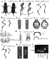
Fig. 1. Cbfβf/fPrx1-Cre mice had dwarfism with shortened limbs
(A) Gross morphology of postnatal 7-day-old (P7) Cbfβf/fPrx1-Cre and wild-type (WT) mice. (B-I) Skeletal analysis by Alizarin red S/Alcian blue staining of P7 Cbfβf/fPrx1-Cre and WT mice. Long bones were shorter (C,D), sutures and fontanelles were widened (black arrow) (F), and ossification of parietal bones (F), frontal bone (F), sternum (H) and hyoid bone (I) was delayed in Cbfβf/fPrx1-Cre mice. Development of ribs (G) and spine (E) was not affected in Cbfβf/fPrx1-Cre mice. (J) Skeletal analysis by Alizarin red S/Alcian blue staining of P30 Cbfβf/fPrx1-Cre and WT mouse limb. (K) Micro-CT analysis of P30 Cbfβf/fPrx1-Cre and WT mouse femur. (L) PCR was used to determine Cbfβ alleles (f/f, f/+, +/+, or deletion) and the presence of Cre.
Fig. 2
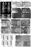
Fig. 2. Cbfβ deficiency resulted in cleidocranial dysplasia-like phenotype in adult mice and skeletal defects in newborn mice
(A) Whole body X-ray of 6-week-old mice showed cleidocranial dysplasia-like phenotype of Cbfβf/fPrx1-Cre mice, including shortened stature, shortened long bones (upper lane, marked by arrowhead), and absent clavicles (lower lane marked by arrowhead and upper lane marked by arrowhead and square). (B) H&E staining of paraffin sections of femurs from newborn Cbfβf/fPrx1-Cre mice and WT mice. (C) Safranin O staining of paraffin sections of femurs from newborn Cbfβf/fPrx1-Cre mice and WT mice. (D) Goldner’s trichrome staining of paraffin sections of femurs from newborn Cbfβf/fPrx1-Cre mice and WT mice. (E) Quantification data of Goldner’s trichrome staining were presented as mean ± SD, n≧6, ns (non-significant), **p<0.01, ***p<0.001 versus WT. (F) TRAP staining of paraffin sections of femurs from newborn Cbfβf/fPrx1-Cre mice and WT mice. P: proliferation zone. R: resting zone. H: hypertrophic zone.
Fig. 3
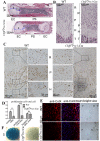
Fig. 3. Cbfβ deficiency retards the development of primary spongiosa and delays chondrocyte proliferation and maturation
(A, B) H&E staining of paraffin sections of femurs from P7 Cbfβf/fPrx1-Cre mice and WT mice. Femur and primary spongiosa were shortened, while the epiphyseal growth plate was elongated in Cbfβf/fPrx1-Cre mice. Columnar structure of proliferative chondrocyte zone was lost in Cbfβf/fPrx1-Cre mice. (C) PCNA staining of paraffin sections of femurs from P7 Cbfβf/fPrx1-Cre mice and WT mice. Second and third columns show magnified images of areas in black boxes. (D) The ratio of proliferating cells in total cells in the three chondrocyte zones of WT and Cbfβf/fPrx1-Cre mice from (C). Data were presented as mean ± SD, n≧6, ns (non-significant), **p<0.01, ***p<0.001 versus the same zone in WT mice. (E) IF staining using frozen sections of P7 mouse femurs showed that ColX expression was decreased in Cbfβf/fPrx1-Cre mice. Bright field views were co-presented on the right panel. (F) Micromass culture of growth plate chondrocytes of newborn mice. PS: primary spongiosa. EC: epiphyseal cartilage. R: resting zone. P: proliferation zone. H: hypertrophic zone. T: trabecular bone.
Fig. 4
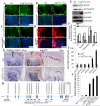
Fig. 4. Expression of Sox9, Ihh, CyclinD1, PTHrP-R, and Cbfβ in chondrocytes of Cbfβf/fPrx1-Cre mice and WT mice
Similar Sox9 expression (A), decreased Ihh expression (B), decreased Cyclin D1 expression (C), and increased PPR expression (D) was observed in femur growth plates of P7 Cbfβf/fPrx1-Cre mice compared to that of WT mice, as detected by IF staining using frozen sections. (E) The expression of Ihh, CyclinD1, PPR and Cbfβ was confirmed by Western blot using protein lysates of femoral cartilage from Cbfβf/fPrx1-Cre and WT newborn mice. (F) Protein levels in (E) were quantified and normalized to tubulin. (G) The expression of Patched (PTC) in the pre-hypertrophic zone of the growth plates in P7 Cbfβf/fPrx1-Cre mice was reduced compared with that in WT mice, as detected by IHC staining using paraffin sections. Third column shows the magnified images of areas in red boxes. Arrowheads indicate positive stains. R: resting zone. P: proliferation zone. H: hypertrophic zone. T: trabecular bone. (H) Schematic display of the Ihh promoter region (−3919/+270). TSS (transcriptional start site), predicted Runx-binding sites and ChIP primer positions were indicated in the figure. (I) ChIP was performed using WT chondrocyte lysates, anti-Cbfβ antibody, and primers as indicated in (H). (J) The Ihh promoter region (−1287/+162) was inserted into the pGL3-basic vector. Primary Cbfβf/fPrx1-Cre chondrocytes were transfected with pGL3-control, pGL3-Ihh+pcDNA3.1a-Cbfβ, or pGL3-Ihh+pcDNA3.1a-Cbfβ+pcNDA3.1a-Runx2. The β-GAL-expression plasmid was also co-transfected. Luciferase activity was detected 48 hours post-transfection, and normalized to β-GAL activity. Data were presented as mean ± SD, n≧6, ns (non-significant), ***p<0.001.
Fig. 5
Fig. 5. Mice lacking Cbfβ had delayed ossification
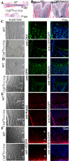
(A,B) H&E staining of the femurs of P30 (postnatal one-month-old) WT and Cbfβf/fPrx1-Cre mice. The femur was shortened and trabecular bone was lost in Cbfβf/fPrx1-Cre mice. The epiphyseal growth plate protruded deep into the diaphysis abnormally (marked by arrowhead). (C-F) IF staining using frozen sections of femurs of P7 (C-E) and newborn (F) WT and Cbfβf/fPrx1-Cre mice. In the Cbfβf/fPrx1-Cre mice, expression of OSX (C), OPN (D) and VEGFA (F) was decreased, but expression of Runx2 (E) was not changed. Bright field views of C-F are co-presented on the left panel. T: trabecular bone. H: hypertrophic zone. P: proliferation zone. pre-H: pre-hypertrophic zone.
Fig. 6
Fig. 6. Cbfβ is required for osteoblastogenesis of calvarial cells
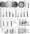
Calvarial cells from Cbfβf/fPrx1-Cre and WT newborn mice were used for primary culture. Osteoblastogenesis was detected by (A) ALP staining on day 14 of osteoblastogenesis and mineralization was detected by (B) Von Kossa staining on day 21 of osteoblastogenesis. (C) Cbfβ expression levels in calvarial cells were detected by qRT-PCR and normalized to Hprt1. (D, E) Expression of Runx2, Opn, Ocn, and Osx in calvarial cells on day 7 (D7) (D) and day 14 (D14) (E) of osteoblastogenesis was detected by qRT-PCR and normalized to Hprt1. (F) Expression of Alp, Ibsp and RANKL in calvarial cells on day 14 of osteoblastogenesis was detected qRT-PCR and normalized to Hprt1. (G) Western blot was applied to detect the protein levels of Runx2, Ocn, and Cbfβ in WT and mutant (MT) calvarial cells on day 7, 14, and 21 (D7, D14 and D21) of osteoblastogenesis. (H) Expression levels of Runx2-I and Runx2-II in calvaria were determined by qRT-PCR and normalized to Gapdh. (I) Western blot was applied to detect the protein levels of Runx1 and Runx3 in WT and mutant calvarial cells on day 7, 14, and 21 of osteoblastogenesis. Results were presented as mean ± SD, n≧6, ns (non-significant), *p<0.05, ***p<0.005.

Similar articles

Cited by

References

    1. Speck NA, Terryl S. A new transcription factor family associated with human leukemias. Crit Rev Eukaryot Gene Expr. 1995;5(3-4):337–64. - PubMed
    1. Levanon D, Negreanu V, Bernstein Y, Bar-Am I, Avivi L, Groner Y. AML1, AML2, and AML3, the human members of the runt domain gene-family: cDNA structure, expression, and chromosomal localization. Genomics. 1994;23(2):425–32. - PubMed
    1. Komori T, Yagi H, Nomura S, Yamaguchi A, Sasaki K, Deguchi K, Shimizu Y, Bronson RT, Gao YH, Inada M, Sato M, Okamoto R, Kitamura Y, Yoshiki S, Kishimoto T. Targeted disruption of Cbfa1 results in a complete lack of bone formation owing to maturational arrest of osteoblasts. Cell. 1997;89(5):755–764. - PubMed
    1. Yoshida CA, Yamamoto H, Fujita T, Furuichi T, Ito K, Inoue K, Yamana K, Zanma A, Takada K, Ito Y, Komori T. Runx2 and Runx3 are essential for chondrocyte maturation, and Runx2 regulates limb growth through induction of Indian hedgehog. Genes Dev. 2004;18(8):952–63. - PMC - PubMed
    1. Kimura A, Inose H, Yano F, Fujita K, Ikeda T, Sato S, Iwasaki M, Jinno T, Ae K, Fukumoto S, Takeuchi Y, Itoh H, Imamura T, Kawaguchi H, Chung UI, Martin JF, Iseki S, Shinomiya K, Takeda S. Runx1 and Runx2 cooperate during sternal morphogenesis. Development. 2010;137(7):1159–67. - PMC - PubMed

Publication types

MeSH terms