. 2014 Nov;29(11):2456-67.
doi: 10.1002/jbmr.2278.

Mechanical load increases in bone formation via a sclerostin-independent pathway

Affiliations

Mechanical load increases in bone formation via a sclerostin-independent pathway

A Morse et al. J Bone Miner Res. 2014 Nov.

Abstract

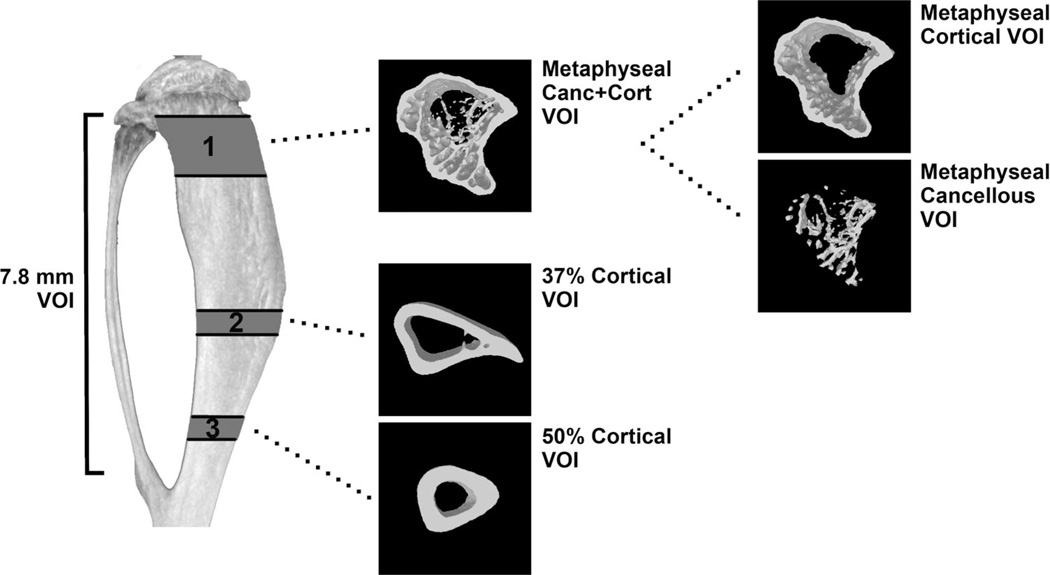
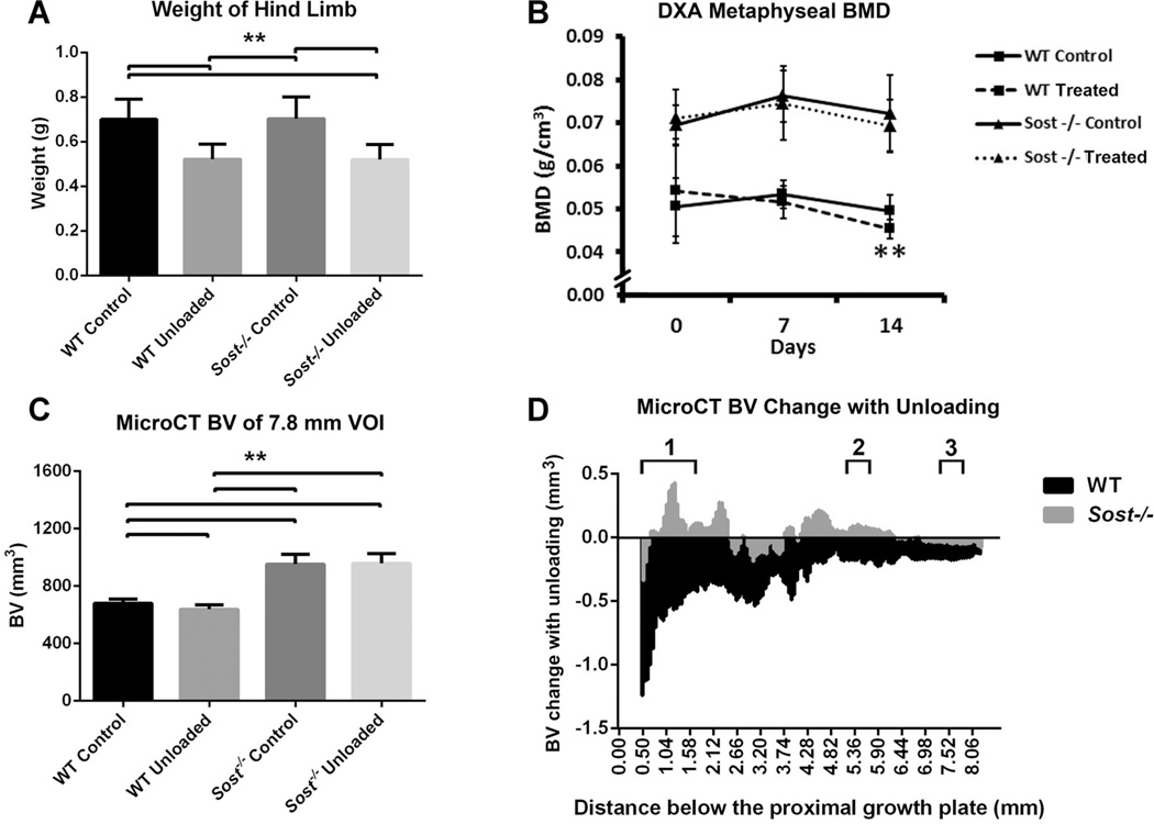
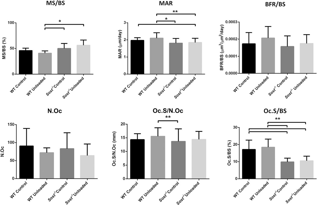
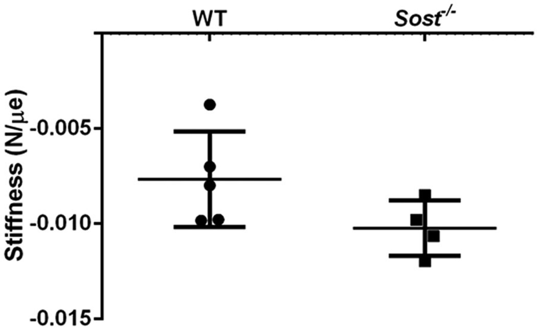
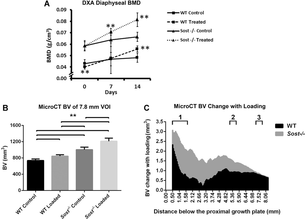
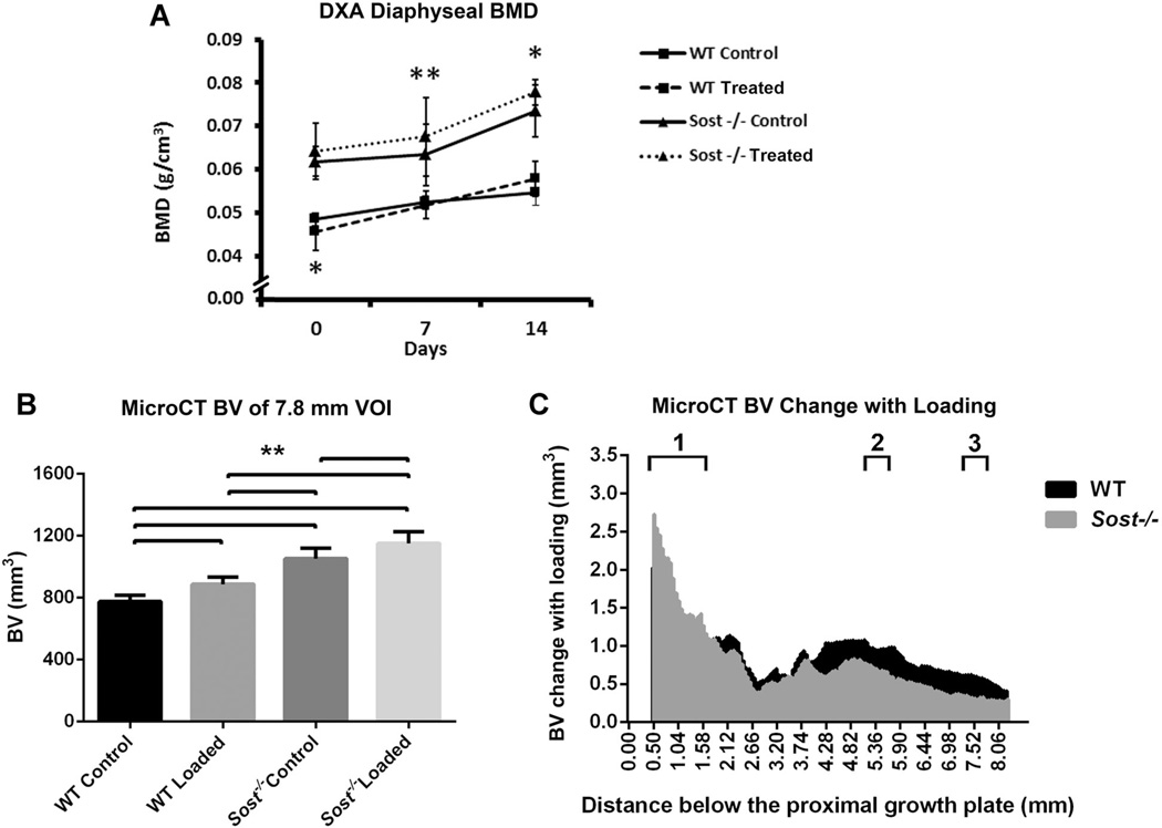
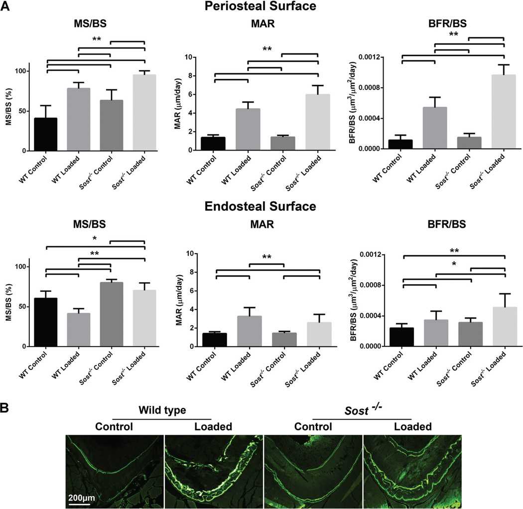
Sclerostin, encoded by the Sost gene, is an important negative regulator of bone formation that has been proposed to have a key role in regulating the response to mechanical loading. To investigate the effect of long-term Sclerostin deficiency on mechanotransduction in bone, we performed experiments on unloaded or loaded tibiae of 10 week old female Sost-/- and wild type mice. Unloading was induced via 0.5U botulinum toxin (BTX) injections into the right quadriceps and calf muscles, causing muscle paralysis and limb disuse. On a separate group of mice, increased loading was performed on the left tibiae through unilateral cyclic axial compression of equivalent strains (+1200 µe) at 1200 cycles/day, 5 days/week. Another cohort of mice receiving equivalent loads (-9.0 N) also were assessed. Contralateral tibiae served as normal load controls. Loaded/unloaded and normal load tibiae were assessed at day 14 for bone volume (BV) and formation changes. Loss of BV was seen in the unloaded tibiae of wild type mice, but BV was not different between normal load and unloaded Sost-/- tibiae. An increase in BV was seen in the loaded tibiae of wild type and Sost-/- mice over their normal load controls. The increased BV was associated with significantly increased mid-shaft periosteal mineralizing surface/bone surface (MS/BS), mineral apposition rate (MAR), and bone formation rate/bone surface (BFR/BS), and endosteal MAR and BFR/BS. Notably, loading induced a greater increase in periosteal MAR and BFR/BS in Sost-/- mice than in wild type controls. Thus, long-term Sclerostin deficiency inhibits the bone loss normally induced with decreased mechanical load, but it can augment the increase in bone formation with increased load.

Keywords: Bone; Loading; Mechanotransduction; Sclerostin; Unloading; WNT.


Figures

Fig. 1
Fig. 1
A 7.8 mm VOI of the tibia starting 0.5 mm below the proximal growth plate was assessed. Sub-regional analysis was performed in 3 VOIs. VOI 1 (Metaphyseal Canc + Cort) represents metaphyseal bone with 1.2 mm height starting 0.5 mm below the growth plate. The Cortical and Cancellous bone compartments of this VOI were analysed separately and together. VOI 2 (37% Cortical) and VOI 3 (50% Cortical) represents diaphyseal cortical bone in two 0.5 mm height VOIs situated 37% and 50% down from the proximal tibia.
Fig. 2
Fig. 2
Control and unloaded tibiae of WT (wild type) and Sost−/− mice: (A) End point weight of control and BTX-treated hind limbs: tibia, fibula, femur and muscle, excluding skin and feet; (B) DXA measured mean bone mineral (BMD) of the tibial metaphysis at days 0, 7, and 14. Bars represent ± SD; (C) MicroCT measured mean bone volume, within the 7.8 mm VOI of the tibiae. Bars represent + SD; (D) Bone volume change between unloaded and control tibiae along the 7.8 mm VOI. 1–3. Localities of sub-regional VOIs: 1. Metaphyseal VOI, 2. 37% Cortical VOI, 3. 50% Cortical VOI. **p < 0.01.
Fig. 3
Fig. 3
Histomorphometric analysis of unloaded and control wild type (WT) and Sost−/− tibiae within metaphyseal cancellous bone. MS/BS, mineralizing surface/bone surface; MAR, mineral apposition rate/day; BFR/BS, bone formation rate/bone surface; N.Oc, osteoclast number; Oc.S/N.Oc, osteoclast surface/osteoclast number; Oc.S/BS, osteoclast surface/bone surface. Bars represent + SD, n = 8–10 per group. **p < 0.01, *p < 0.05.
Fig. 4
Fig. 4
Mean stiffness of wild type (WT) and Sost−/− tibiae measured by strain gauging of the mid-diaphysis. Sost−/− tibiae trended toward being stiffer than WT controls (p = 0.09). Bars represent ± SD, n = 4–5 per group.
Fig. 5
Fig. 5
Control and strain-matched (1200µe) loaded tibiae of WT (wild type) and Sost−/− mice: (A) DXA measured mean bone mineral (BMD) of the tibial diaphysis at days 0, 7, and 14. Bars represent ± SD; (B) MicroCT measured mean bone volume, within the 7.8 mm VOI of the tibiae. Bars represent + SD; (C) Bone volume change between loaded and control tibiae along the 7.8 mm VOI. 1–3. Localities of sub-regional VOIs: 1. Metaphyseal VOI, 2. 37% Cortical VOI, 3. 50% Cortical VOI. **p < 0.01
Fig. 6
Fig. 6
Control and load-matched (−9.0N) loaded tibiae of WT (wild type) and Sost−/− mice: (A) DXA measured mean bone mineral (BMD) of the tibial diaphysis at days 0, 7, and 14. Bars represent ± SD; (B) MicroCT measured mean bone volume, within the 7.8 mm VOI of the tibiae. Bars represent + SD; (C) Bone volume change between loaded and control tibiae along the 7.8 mm VOI. 1–3. Localities of sub-regional VOIs: 1. Metaphyseal VOI, 2. 37% Cortical VOI, 3. 50% Cortical VOI. **p < 0.01, *p < 0.05.
Fig. 7
Fig. 7
Histomorphometric analysis of strain-matched (1200µe) loaded and control wild type (WT) and Sost−/− tibiae within the diaphyseal 37% Cortical VOI. (A) Periosteal and endosteal surfaces were analysed for: MS/BS, mineralizing surface/bone surface; MAR mineral apposition rate/day; BFR/BS, bone formation rate/bone surface. Bars represent + SD, n = 8–10 per group. **p < 0.01, *p < 0.05. (B) Representative images of Calcein bone labeling.

References

    1. Poole KE, van Bezooijen RL, Loveridge N, et al. Sclerostin is a delayed secreted product of osteocytes that inhibits bone formation. FASEB J. 2005;19(13):1842–1844. - PubMed
    1. van Bezooijen RL, ten Dijke P, Papapoulos SE, Lowik CW. SOST/sclerostin, an osteocyte-derived negative regulator of bone formation. Cytokine Growth Factor Rev. 2005;16(3):319–327. - PubMed
    1. Semenov M, Tamai K, He X. SOST is a ligand for LRP5/LRP6 and a Wnt signaling inhibitor. J Biol Chem. 2005;280(29):26770–26775. - PubMed
    1. van Bezooijen RL, Bronckers AL, Gortzak RA, et al. Sclerostin in mineralized matrices and van Buchem disease. J Dental Res. 2009;88(6):569–574. - PubMed
    1. Jager A, Gotz W, Lossdorfer S, Rath-Deschner B. Localization of SOST/sclerostin in cementocytes in vivo and in mineralizing periodontal ligament cells in vitro. J Periodontal Res. 2010;45(2):246–254. - PubMed

Publication types

MeSH terms

Substances