

Behçet disease-associated MHC class I residues implicate antigen binding and regulation of cell-mediated cytotoxicity
- PMID: 24821759
- PMCID: PMC4066484
- DOI: 10.1073/pnas.1406575111
Behçet disease-associated MHC class I residues implicate antigen binding and regulation of cell-mediated cytotoxicity
Abstract
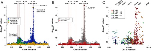
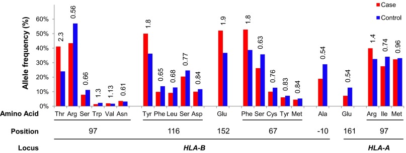
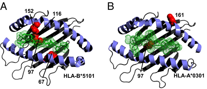
The HLA protein, HLA-B*51, encoded by HLA-B in MHC, is the strongest known genetic risk factor for Behçet disease (BD). Associations between BD and other factors within the MHC have been reported also, although strong regional linkage disequilibrium complicates their confident disentanglement from HLA-B*51. In the current study, we examined a combination of directly obtained and imputed MHC-region SNPs, directly obtained HLA-B locus types, and imputed classical HLA types with their corresponding polymorphic amino acid residues for association with BD in 1,190 cases and 1,257 controls. SNP mapping with logistic regression of the MHC identified the HLA-B/MICA region and the region between HLA-F and HLA-A as independently associated with BD (P < 1.7 × 10(-8)). HLA-B*51, -A*03, -B*15, -B*27, -B*49, -B*57, and -A*26 each contributed independently to BD risk. We directly examined rs116799036, a noncoding SNP upstream of HLA-B that was recently suggested to underlie the association of HLA-B*51 with BD, but we were unable to replicate that finding in our collection. Instead, we mapped the BD association to seven MHC class I (MHC-I) amino acid residues, including anchor residues that critically define the selection and binding of peptides to MHC-I molecules, residues known to influence MHC-I-killer immunoglobulin-like receptor interactions, and a residue located in the signal peptide of HLA-B. The locations of these variants collectively implicate MHC-I peptide binding in the pathophysiology of BD. Furthermore, several lines of evidence suggest a role for altered regulation of cellular cytotoxicity in BD pathogenesis.
Keywords: HLA imputation; antigen presentation; autoinflammation; killer immunoglobulin-like receptors; natural killer cells.
Conflict of interest statement
The authors declare no conflict of interest.
Figures
Comment in
-
HLA-B*51 the primary risk in Behçet disease.Proc Natl Acad Sci U S A. 2014 Jun 17;111(24):8706-7. doi: 10.1073/pnas.1407307111. Epub 2014 May 29. Proc Natl Acad Sci U S A. 2014. PMID: 24876276 Free PMC article. No abstract available.
References
-
- Sakane T, Takeno M, Suzuki N, Inaba G. Behçet’s disease. N Engl J Med. 1999;341(17):1284–1291. - PubMed
-
- Mizuki N, et al. Genome-wide association studies identify IL23R-IL12RB2 and IL10 as Behçet’s disease susceptibility loci. Nat Genet. 2010;42(8):703–706. - PubMed
-
- Ono S, Aoki K, Sugiura S, Nakayama E, Itakura K. Letter: HL-A5 and Behçet’s disease. Lancet. 1973;2(7842):1383–1384. - PubMed
-
- Ohno S, et al. Close association of HLA-Bw51 with Behçet’s disease. Arch Ophthalmol. 1982;100(9):1455–1458. - PubMed
Publication types
MeSH terms
Substances
Grants and funding
LinkOut - more resources
Full Text Sources
Other Literature Sources
Medical
Research Materials
