Clinical Trial
. 2014 Jun 24;111(25):E2567-75.
doi: 10.1073/pnas.1406974111. Epub 2014 May 12.

Redemption of autoantibodies on anergic B cells by variable-region glycosylation and mutation away from self-reactivity

Affiliations
Clinical Trial

Redemption of autoantibodies on anergic B cells by variable-region glycosylation and mutation away from self-reactivity

Zahra Sabouri et al. Proc Natl Acad Sci U S A. .

Abstract

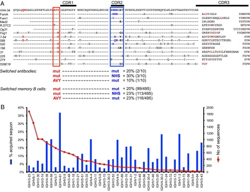
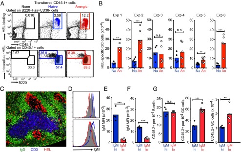
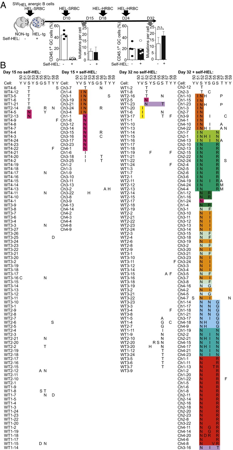
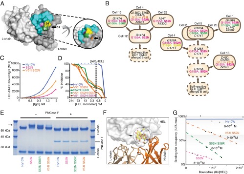
The best-understood mechanisms for achieving antibody self/non-self discrimination discard self-reactive antibodies before they can be tested for binding microbial antigens, potentially creating holes in the repertoire. Here we provide evidence for a complementary mechanism: retaining autoantibodies in the repertoire displayed as low levels of IgM and high IgD on anergic B cells, masking a varying proportion of autoantibody-binding sites with carbohydrates, and removing their self-reactivity by somatic hypermutation and selection in germinal centers (GCs). Analysis of human antibody sequences by deep sequencing of isotype-switched memory B cells or in IgG antibodies elicited against allogeneic RhD+ erythrocytes, vaccinia virus, rotavirus, or tetanus toxoid provides evidence for reactivation of anergic IgM(low) IgD+ IGHV4-34+ B cells and removal of cold agglutinin self-reactivity by hypermutation, often accompanied by mutations that inactivated an N-linked glycosylation sequon in complementarity-determining region 2 (CDR2). In a Hy10 antibody transgenic model where anergic B cells respond to a biophysically defined lysozyme epitope displayed on both foreign and self-antigens, cell transfers revealed that anergic IgM(low) IgD+ B cells form twice as many GC progeny as naïve IgM(hi) IgD+ counterparts. Their GC progeny were rapidly selected for CDR2 mutations that blocked 72% of antigen-binding sites with N-linked glycan, decreased affinity 100-fold, and then cleared the binding sites of blocking glycan. These results provide evidence for a mechanism to acquire self/non-self discrimination by somatic mutation away from self-reactivity, and reveal how varying the efficiency of N-glycosylation provides a mechanism to modulate antibody avidity.

Keywords: affinity maturation; autoimmunity; clonal selection; self-tolerance.


Conflict of interest statement

The authors declare no conflict of interest.

Figures

Fig. 1.
Fig. 1.
Evidence for mutation away from self-reactivity in IGHV4-34 antibodies. (A) Antibodies using IGHV4-34 from normal donors. The germ-line IGHV-34*01 amino acid sequence is shown at the top. In red are the residues of the hydrophobic patch that cause binding to self-antigens on the surface of B cells and erythrocytes, notably I/i carbohydrates, with the AVY sequence boxed. In blue and boxed is the germ-line NHS N-glycosylation sequon in CDR2. Sequons flanking residues that may modulate glycosylation efficiency analogous to Hy10 are also shown in bold. Aligned beneath are the corresponding sequences of specific antibodies (switched IgG antibodies are italicized) elicited by immunization with a foreign antigen, revealed by an IGHV-34*01 Blastn search of the NCBI nonredundant nucleotide database and analyzed using IMGT/V-QUEST. Identity to germ line is denoted by a dash, and substituted residues in CDR3 are in dark red. The percentage of switched antibodies with mutations that inactivate the hydrophobic patch AVY sequence or the core NHS glycosylation sequon, or both, is shown below for the switched antibodies of known specificity and in massively parallel cDNA sequences from memory B cells. Antibody specificities and GenBank accession numbers are as follows: anti-RhD (16): FomA (X64153), Fom1 (X64152), Mad2 (X64159), R.D7C2 (A49385), Og31 (X64156), Fog1 (X64150); anti-rotavirus (17): 7-94 (AF453121); anti-vaccinia (18): 589 (HQ378397), 520 (HQ378388), 166 (HQ378337), 31 (HQ378326), 225 (HQ378353), 274 (HQ378366); and anti-tetanus toxoid (19): 529E18 (JN111017). (B) Repertoire of mutated IGH cDNAs in IgD− CD27+ memory B cells in blood of normal donors, analyzed by 454 massively parallel sequencing, showing the number of sequences for each IGHV gene and the percentage that have acquired an N-glycosylation sequon. Excluded are IGHV genes with germ-line sequons (IGHV4-34, IGHV1-8, IGHV5-a) or with less than 20 reads.
Fig. 2.
Fig. 2.
IgMlow anergic B cells paradoxically make more GC progeny. (AC) CD45.1-marked HEL-specific B cells (105) that were anergic (An) or naïve (Na) were injected into normal C57BL/6 recipient mice together with 108 HEL-SRBCs or HEL2X-SRBCs. (A) Representative flow cytometric analysis of recipient spleen cells 5 d after immunization with HEL2X-SRBCs, gating on Fas+ CD38− GC B cells to enumerate percentage CD45.1+ donor-derived (Upper) or gating on CD45.1+ donor B cells (Lower) and measuring the percentage differentiated into B220low plasma cells with high intracellular HEL-binding antibody or into GC cells. (B) Arithmetic means and data from individual recipients immunized with HEL2X-SRBCs (exp 1–4) or HEL-SRBCs (exp 5 and 6). Statistical analysis was by t test: **P ≤ 0.01, ***P ≤ 0.001; n.s., not significant. (C) Representative immunofluorescence staining of a spleen cryosection from the recipient of anergic cells on day 5 after immunization with HEL2X-SRBCs, locating most HEL-binding progeny (red) in GCs. (DG) Mature CD23+ B cells from C57BL/6 mice were stained with F(ab′) anti-IgM, and cells in the lowest (IgMlo; red histograms) or highest (IgMhi; blue histograms) quartile of cell-surface IgM were sorted and injected into B6.SJL-CD45.2 congenic recipients, and the recipients were immunized with SRBCs. (D) Distribution of surface IgM on the sorted populations compared with unsorted CD23+ B cells (gray-filled histogram) on day 0 (Upper) and compared with CD45.1+ recipient B220+ B cells 6 d after transfer (Lower). (E and F) Surface IgM mean fluorescence intensity (MFI) on the sorted populations from separate donors at the time of transfer (E) and in separate recipients after 6 d (F). (G) Frequency of CD45.2+ donor-derived cells among B220+ Fas− GL7− B cells (Left) or among B220+ Fas+ GL7+ GC cells (Center) in individual recipients, and the total number of donor-derived GC cells (Right). Statistical analysis was done as above.
Fig. 3.
Fig. 3.
Recurring VH CDR2 mutations selected by self-antigen. (A) Anergic CD45.1-marked HEL-specific B cells (105) from SWHEL x ML5 donor mice were transferred into non-Tg (no self-HEL) or ML5 HEL-Tg (+self-HEL) C57BL/6 recipients together with HEL-SRBCs, and recipients were boosted on the indicated days with HEL-SRBCs or HEL-HRBCs. The % CD45.1+ cells among B220+ Fas+ GL7+ GC B cells in individual recipients on day 15 or 32 is shown. Single CD45.1+ B220+ Fas+ GL7+ cells were sorted into microtiter plates for Hy10 VDJH exon sequencing, yielding the mean number of VDJH nucleotide substitutions in B cells from individual recipients and the SD of this mean between recipients. (B) Amino acid changes in CDR2 in single sorted B cells after 15 or 32 d in recipients with or without self-HEL. Cells from different mice are denoted by the prefix Ch1–Ch4 for the four HEL-transgenic mice on day 15 and day 32, and WT1–4 for the four non-Tg mice at each time point. From each of the mice analyzed, the individual cells are numbered with a suffix from 1 to 25. The Hy10 sequence for CDR2 residues 50–59 is shown above. Substitutions in individual cells are shown, clustered by sequence similarity. The different colors denote recurring combinations of CDR2 mutations, often found in several independent recipient mice, as indicated by the Ch1–Ch4 prefix.
Fig. 4.
Fig. 4.
Mutation away from self-reactivity by modulating glycosylation, affinity, and avidity. (A) Location of mutated VH residues relative to the H-chain contact surface with lysozyme in blue, from Protein Data Bank (PDB) ID code 3D9A (32). (B) Representative GC B-cell genealogies in individual recipients on day 15 (Left; from mouse number Ch3) and day 32 (Right; from mouse number Ch1). Clonally related cells were identified by shared silent mutations in nonhotspot VH nucleotides (italicized black). Recurrent nonsynonymous nucleotide and corresponding amino acid substitutions are colored; other mutations are shown in black. Sequenced cells are numbered as in Fig. 2, and inferred precursors are indicated by dashed circles. (C and D) IgG1 antibodies with the wild-type H-chain sequence (Hy10W) or the indicated CDR2 substitutions were expressed in CHO cells, and binding to HEL-SRBCs was detected by fluorescent anti-IgG antibody and flow cytometry. MFI, geometric mean fluorescence intensity of IgG fluorescence on gated single erythrocytes. (C) Binding in the absence of competing soluble HEL. (D) Percent inhibition of binding to HEL-SRBCs of 0.5 nM IgG in the presence of varying concentrations of HEL monomer in solution. The dashed line denotes the mean concentration of self-HEL in the extracellular fluid of ML5 mice. (E) Hy10 IgG1 antibodies with the wild-type H-chain sequence (Hy10W) or the indicated CDR2 substitutions were expressed in 293T cells, purified, and analyzed by SDS/PAGE under reducing conditions and stained with Coomassie blue. Where indicated, N-linked carbohydrates were removed from the antibodies by treatment with PNGase F (+). The arrow indicates mobility shift due to VH glycosylation. (F) Molecular model of V-region glycosylation, with carbohydrate (arrow) occupying the HEL antigen-binding site. The model was generated using the structure of Hy10W bound to HEL (cartoon and transparent surface; PDB ID code 3D9A), with a single N-linked branched NAG-FUC-NAG trisaccharide modeled onto Kabat position 52 of the variable heavy domain (colored sticks; glycan-derived from PDB ID code 3CA1). (G) Biolayer interferometry measurements of binding of HEL antigen in solution to immobilized Hy10 IgG antibodies. Plotted is binding site occupancy (% Hy10W maximum) versus Octet IU intensity units per antigen concentration (nm/M).

Comment in

  • Redemption of autoreactive B cells.
    Haynes BF, Verkoczy L, Kelsoe G. Haynes BF, et al. Proc Natl Acad Sci U S A. 2014 Jun 24;111(25):9022-3. doi: 10.1073/pnas.1407877111. Epub 2014 Jun 11. Proc Natl Acad Sci U S A. 2014. PMID: 24920593 Free PMC article. No abstract available.

References

    1. Burnet FM. The Clonal Selection Theory of Acquired Immunity. Cambridge, UK: Cambridge Univ Press; 1959.
    1. Goodnow CC, Ohashi PS. Immunological tolerance. In: Paul WE, editor. Fundamental Immunology. 7th Ed. Philadelphia: Wolters Kluwer Health/Lippincott Williams & Wilkins; 2013. pp. 765–794.
    1. Jerne NK. The somatic generation of immune recognition. Eur J Immunol. 1971;1(1):1–9. - PubMed
    1. Diaz M, Klinman NR. Relative roles of somatic and Darwinian evolution in shaping the antibody response. Immunol Res. 2000;21(2-3):89–102. - PubMed
    1. Goodnow CC, Crosbie J, Jorgensen H, Brink RA, Basten A. Induction of self-tolerance in mature peripheral B lymphocytes. Nature. 1989;342(6248):385–391. - PubMed

Publication types

MeSH terms