. 2014 May 27;111(21):7753-8.
doi: 10.1073/pnas.1401857111. Epub 2014 May 12.

RIP1 suppresses innate immune necrotic as well as apoptotic cell death during mammalian parturition

Affiliations

RIP1 suppresses innate immune necrotic as well as apoptotic cell death during mammalian parturition

William J Kaiser et al. Proc Natl Acad Sci U S A. .

Abstract

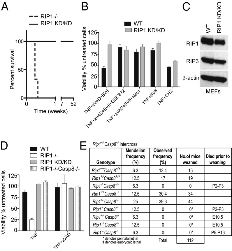
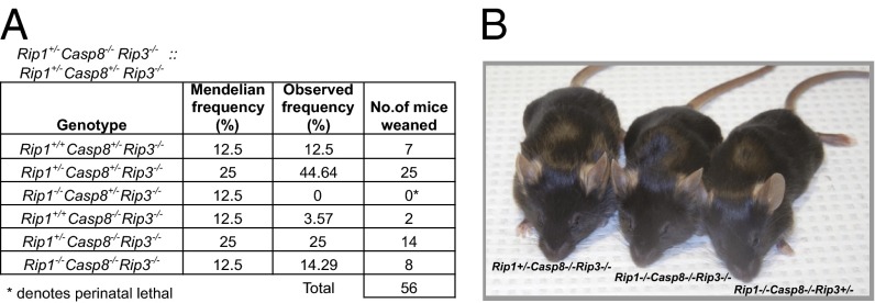
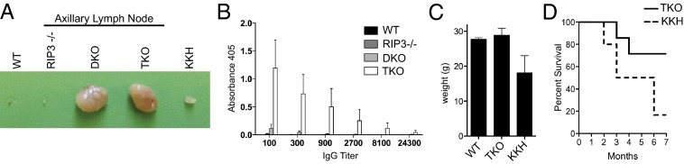
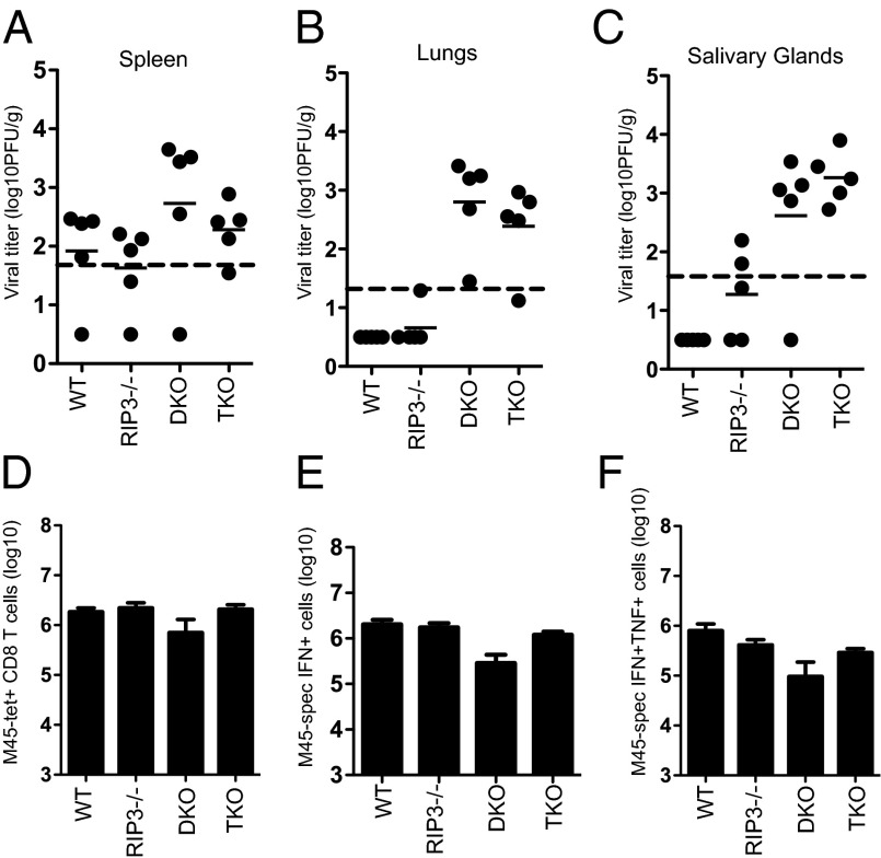
The pronecrotic kinase, receptor interacting protein (RIP1, also called RIPK1) mediates programmed necrosis and, together with its partner, RIP3 (RIPK3), drives midgestational death of caspase 8 (Casp8)-deficient embryos. RIP1 controls a second vital step in mammalian development immediately after birth, the mechanism of which remains unresolved. Rip1(-/-) mice display perinatal lethality, accompanied by gross immune system abnormalities. Here we show that RIP1 K45A (kinase dead) knockin mice develop normally into adulthood, indicating that development does not require RIP1 kinase activity. In the face of complete RIP1 deficiency, cells develop sensitivity to RIP3-mixed lineage kinase domain-like-mediated necroptosis as well as to Casp8-mediated apoptosis activated by diverse innate immune stimuli (e.g., TNF, IFN, double-stranded RNA). When either RIP3 or Casp8 is disrupted in combination with RIP1, the resulting double knockout mice exhibit slightly prolonged survival over RIP1-deficient animals. Surprisingly, triple knockout mice with combined RIP1, RIP3, and Casp8 deficiency develop into viable and fertile adults, with the capacity to produce normal levels of myeloid and lymphoid lineage cells. Despite the combined deficiency, these mice sustain a functional immune system that responds robustly to viral challenge. A single allele of Rip3 is tolerated in Rip1(-/-)Casp8(-/-)Rip3(+/-) mice, contrasting the need to eliminate both alleles of either Rip1 or Rip3 to rescue midgestational death of Casp8-deficient mice. These observations reveal a vital kinase-independent role for RIP1 in preventing pronecrotic as well as proapoptotic signaling events associated with life-threatening innate immune activation at the time of mammalian parturition.

Keywords: MLKL; herpesvirus; interferon.


Conflict of interest statement

Conflict of interest statement: P.J.G., J.B., and S.B.B. are employees of GlaxoSmithKline.

Figures

Fig. 1.
Fig. 1.
Survival of Rip1KD/KD but not Rip1−/−Casp8−/− mice implicates programmed necrosis in perinatal death of Rip1−/− mice. (A) Kaplan–Meier survival plots of Rip1KD/KD and Rip1−/− mice. (B) Viability of WT and Rip1KD/KD MEFs by Cell Titer-Glo (Promega) assay (10), determined 12 h after stimulation with necrotic or apoptotic stimuli. Necroptosis was induced by treatment with TNF (25 ng/mL) in the presence of zVAD-fmk (zVAD, 25 µM) and BV6 (1 µM) with or without inhibitors GSK’872 (3 µM) or Nec-1 (30 µM). Apoptosis was induced by treatment with TNF in the presence of cyclohexamide (5 µg/mL). (C) Immunoblot of RIP1, RIP3, and β-actin levels in WT and RIP1KD/KD MEFs. (D) Viability of indicated genotypes of primary MEFs at 18 h after treatment with TNF in the presence or absence of zVAD-fmk. (E) Epistatic analysis of mice born after intercross of Rip1+/−Casp8+/− mice, with the day of embryonic (E) or perinatal (P) death before weaning indicated in the last column.
Fig. 2.
Fig. 2.
Rip1−/− and Rip1−/−Casp8−/− fibroblasts exhibit sensitivity to innate immune signaling death. (A) Photomicrographs of SV40 immortalized WT and Rip1−/− fibroblasts treated with IFNβ (5 ng/mL), IFNγ (5 ng/mL), TNF (50 ng/mL), or cytosolic poly(I:C) (2 μg/mL transfected in 6 μL Lipofectamine 2000) for 48 h. (B) Time course of viability of immortalized WT and Rip1−/− fibroblasts treated with IFNγ, IFNβ, TNF, or poly(I:C). (Inset) Immunoblot of RIP1 and β-actin levels in immortalized WT and Rip1−/− fibroblasts. (C) Viability of MEFs with the indicated genotypes at 48 h posttreatment with IFNβ (5 ng/mL). (D) Immunoblot of MLKL and RIP3 levels in Rip1−/−Casp8−/− MEFs transfected with nontargeting (NT), RIP3, or MLKL siRNA. (E) Viability assay of Rip1−/−Casp8−/− MEFs 48 h posttransfection with NT, RIP3, or MLKL siRNA treated with IFNβ (5 ng/mL) for 48 h. (F) Viability assay of Rip1−/−Casp8−/− MEFs in the presence or absence of zVAD-fmk (25 µM), GSK’872 (1, 3, or 5 µM) at 60 h posttreatment. Viability was determined by Cell Titer-Glo assay.
Fig. 3.
Fig. 3.
Rip1−/−Casp8−/−Rip3−/− and the Rip1−/−Casp8−/−Rip3+/− mice are viable. (A) Epistatic analysis of mice born following Rip1+/−Casp8+/−Rip3−/− intercross. (B) Image of 5-wk-old TKO, KKH, and Rip1+/−Casp8−/−RIP3−/− mice.
Fig. 4.
Fig. 4.
Immune phenotype of Rip1−/−Casp8−/−Rip3−/− and Rip1−/−Casp8−/−Rip3+/− mice. (A) Axillary lymph nodes from WT, Rip3−/−, DKO, TKO, and KKH mice. (B) Relative serum levels of double-stranded (ds) DNA-specific antibodies measured by ELISA in WT, Rip3−/−, DKO, and TKO mice. (C) Weights of adult WT, TKO, and KKH mice. (D) Kaplan–Meier survival plots comparing survival of TKO and KKH mice through 7 mo of age.
Fig. 5.
Fig. 5.
Rip1−/−Casp8−/−Rip3−/− mice retain the ability to mount an adaptive immune response to virus infection. (AC) MCMV titers in spleen (A), lung (B), and salivary glands (C) from 12- to 16-wk-old WT, Rip3−/−, DKO, or TKO mice 7 d postinoculation with 106 pfu virus. Dashed line indicates limit of detection for each organ type. Shown is log titer of virus per gram of tissue from indvidual mice (five mice per group). (D) Total number of CD8 T cells in spleen recognized by M45-specific MHC class I tetramer in WT, Rip3−/−, DKO, or TKO mice 7 d postinfection. (E) Frequency of splenic CD8 T cells producing IFNγ when stimulated with M45 peptide. (F) Frequency of splenic CD8 T cells producing both IFNγ and TNF when stimulated with M45 peptide.

References

    1. Green DR, Oberst A, Dillon CP, Weinlich R, Salvesen GS. RIPK-dependent necrosis and its regulation by caspases: A mystery in five acts. Mol Cell. 2011;44(1):9–16. - PMC - PubMed
    1. Feoktistova M, Geserick P, Panayotova-Dimitrova D, Leverkus M. Pick your poison: The Ripoptosome, a cell death platform regulating apoptosis and necroptosis. Cell Cycle. 2012;11(3):460–467. - PubMed
    1. Kaiser WJ, Upton JW, Mocarski ES. Viral modulation of programmed necrosis. Curr Opin Virol. 2013;3(3):296–306. - PMC - PubMed
    1. Mocarski ES, Kaiser WJ, Livingston-Rosanoff D, Upton JW, Daley-Bauer LP. True grit: Programmed necrosis in antiviral host defense, inflammation, and immunogenicity. J Immunol. 2014;192(5):2019–2026. - PMC - PubMed
    1. Kelliher MA, et al. The death domain kinase RIP mediates the TNF-induced NF-kappaB signal. Immunity. 1998;8(3):297–303. - PubMed

Publication types

MeSH terms