. 2014 May 27;111(21):7630-5.
doi: 10.1073/pnas.1403779111. Epub 2014 May 12.

The linker connecting the two kringles plays a key role in prothrombin activation

Affiliations

The linker connecting the two kringles plays a key role in prothrombin activation

Nicola Pozzi et al. Proc Natl Acad Sci U S A. .

Abstract

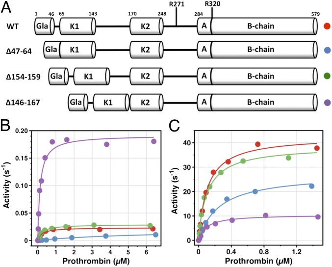
The zymogen prothrombin is proteolytically converted by factor Xa to the active protease thrombin in a reaction that is accelerated >3,000-fold by cofactor Va. This physiologically important effect is paradigmatic of analogous cofactor-dependent reactions in the coagulation and complement cascades, but its structural determinants remain poorly understood. Prothrombin has three linkers connecting the N-terminal Gla domain to kringle-1 (Lnk1), the two kringles (Lnk2), and kringle-2 to the C-terminal protease domain (Lnk3). Recent developments indicate that the linkers, and particularly Lnk2, confer on the zymogen significant flexibility in solution and enable prothrombin to sample alternative conformations. The role of this flexibility in the context of prothrombin activation was tested with several deletions. Removal of Lnk2 in almost its entirety (ProTΔ146-167) drastically reduces the enhancement of thrombin generation by cofactor Va from >3,000-fold to 60-fold because of a significant increase in the rate of activation in the absence of cofactor. Deletion of Lnk2 mimics the action of cofactor Va and offers insights into how prothrombin is activated at the molecular level. The crystal structure of ProTΔ146-167 reveals a contorted architecture where the domains are not vertically stacked, kringle-1 comes within 9 Å of the protease domain, and the Gla-domain primed for membrane binding comes in contact with kringle-2. These findings broaden our molecular understanding of a key reaction of the blood coagulation cascade where cofactor Va enhances activation of prothrombin by factor Xa by compressing Lnk2 and morphing prothrombin into a conformation similar to the structure of ProTΔ146-167.

Keywords: clotting factor; structural biology; zymogen activation.


Conflict of interest statement

The authors declare no conflict of interest.

Figures

Fig. 1.
Fig. 1.
(A) Schematic representation of the structural domains of prothrombin and the deletion mutants characterized in this study. (B and C) Generation of activity in the absence (B) or presence (C) of cofactor Va for wild-type and mutant prothrombins. Experimental data obey Michaelis–Menten kinetics with best-fit parameter values reported in Table 1. Experimental conditions are as follows: 150 mM NaCl, 5 mM CaCl2, 0.1% PEG 8000, and 20 mM Tris at pH 7.4 and 25 °C. Activity on the y axis is expressed in s−1 units to facilitate visualization of kcat values. Colors refer to the constructs in A: wt (red), proTΔ47–64 (blue), ProTΔ154–159 (green), ProTΔ146–167 (purple).
Fig. 2.
Fig. 2.
Activation of prothrombin wild-type and mutants in the absence (-FVa) or presence (+FVa) of cofactor Va. In the absence of cofactor Va, activation of prothrombin wild-type and mutants leads to accumulation of prethrombin-2 (Pre-2) and fragment 1.2. (F1.2) as main products. Notably, the band of the intact ProTΔ146–167 disappears at a rate considerably faster than wild-type prothrombin (see also Fig. 1). The inhibitor DAPA was added to inhibit thrombin and meizothrombin activity. Nonetheless, cleavage at R155 and formation of prethrombin-1 (Pre-1) was detected in prothrombin wild-type and ProTΔ47–64, but not for ProTΔ146–167 and ProTΔ154–159 where R155 is missing. The cleavage is due to factor Xa and not thrombin because the prethrombin-1 band is also observed when the prothrombin mutant S525A is used as substrate to generate a catalytically inactive product. The chemical identity of this band was verified by N-terminal sequencing (156SEGSS160). The faint bands around 30 kDa refer to fragment 1 because they are not detected when R155 is missing. In the presence of cofactor Va, prothrombin wild-type is cleaved at R320 along the meizothrombin pathway to generate fragment F1.2.A and the B chain. The same profile is observed for ProTΔ154–159 and ProTΔ146–167. However, activation of ProTΔ47–64 clearly differs from wild type and follows the prethrombin-2 pathway. Experimental conditions are 150 mM NaCl, 5 mM CaCl2, 0.1% PEG 8000, and 20 mM Tris at pH 7.4 and 25 °C. Time points for the SDS/PAGE experiments are in minutes.
Fig. 3.
Fig. 3.
(A) X-ray crystal structure of ProTΔ146–167 in the Ca2+-bound form showing the arrangement of the various domains (Ac, A chain; Bc, B chain; GD, Gla domain; K1, kringle-1; K2, kringle-2) that are not vertically stacked. The structure spans approximately 87 Å in length, from the tip of F4 in the Gla domain to the site of cleavage R320 that is fully exposed to solvent (see also Fig. 4). The site of cleavage at R271 is in a disordered region in Lnk3. The active site region is circled. Details of the Gla domain containing five bound Ca2+ ions are shown in Fig. 4. (B) Overlay of the structures of ProTΔ146–167 (ribbon colored as in A) and GD-ProT (wheat) reported recently (15). The two structures (rmsd = 1.1 Å) overlap significantly at the level of kringle-2 and the protease domain (rmsd = 0.77 Å), but differ sharply in the orientation of kringle-1 that shifts >50 Å in ProTΔ146–167 from its position in GD-ProT (K1 labeled in purple).
Fig. 4.
Fig. 4.
(A) Artificial connection between Q145 in kringle-1 and E168 in kringle-2 generated by deletion of Lnk2 in ProTΔ146–167. (B) Activation domain of ProTΔ146–167 showing the side chain of R320 at the site of cleavage fully exposed to solvent for proteolytic attack. (C) Active-site metrics of ProTΔ146–167 showing the Cα-Cα distances G548–G523 (G216–G193) and S525–D519 (S195–D189) measuring the width and depth of the catalytic pocket (chymotrypsin numbering is shown by parentheses). W468 (W148) from the autolysis loop penetrates the active site pushing away the flipped W547 (W215) and W370 (W60d) in the 60-loop and pulling in the 547–549 (215–217) segment. Electron densities in AC are 2 F0–Fc maps contoured at 1 σ. (D) Architecture of the Gla domain of ProTΔ146–167 revealing four helical segments N12-T21 (H1), S23-L31 (H2), T37-A50 (H3), P53-G63 (H4) and five Ca2+ ions bound to sites 2–6 in the Tulinsky’s numbering (24). (E) Detailed view of the five Ca2+ ions bound to γ-carboxyglutamic acid residues (labeled as E) of the Gla domain organizing the hydrophobic ω-loop (F4-L5-V8) for membrane binding. Ca2+ binding sites (numbered 2–6 from right to left, see also C) were validated with the program VALE (39) that returned valence units/coordination of 1.7/5 (site 2), 1.1/4 (site 3), 1.4/8 (site 4), 1.0/6 (site 5), and 0.64/2 (site 6). The low numbers for site 6 suggest weak binding affinity for this site and contribution of water molecules to the coordination shell not detected because of the low resolution. Electron density is an F0–Fc map contoured at 3.5 σ and calculated after removing the bound Ca2+ ions.

References

    1. Rosing J, Tans G, Govers-Riemslag JW, Zwaal RF, Hemker HC. The role of phospholipids and factor Va in the prothrombinase complex. J Biol Chem. 1980;255(1):274–283. - PubMed
    1. Nesheim ME, Taswell JB, Mann KG. The contribution of bovine Factor V and Factor Va to the activity of prothrombinase. J Biol Chem. 1979;254(21):10952–10962. - PubMed
    1. Weinreb GE, Mukhopadhyay K, Majumder R, Lentz BR. Cooperative roles of factor V(a) and phosphatidylserine-containing membranes as cofactors in prothrombin activation. J Biol Chem. 2003;278(8):5679–5684. - PubMed
    1. Wu JR, et al. Role of procoagulant lipids in human prothrombin activation. 1. Prothrombin activation by factor X(a) in the absence of factor V(a) and in the absence and presence of membranes. Biochemistry. 2002;41(3):935–949. - PubMed
    1. Krem MM, Di Cera E. Evolution of enzyme cascades from embryonic development to blood coagulation. Trends Biochem Sci. 2002;27(2):67–74. - PubMed

Publication types

MeSH terms

LinkOut - more resources