. 2014 May 12;205(3):313-24.
doi: 10.1083/jcb.201312024.

Pericentromere tension is self-regulated by spindle structure in metaphase

Affiliations

Pericentromere tension is self-regulated by spindle structure in metaphase

Jeremy M Chacón et al. J Cell Biol. .

Abstract

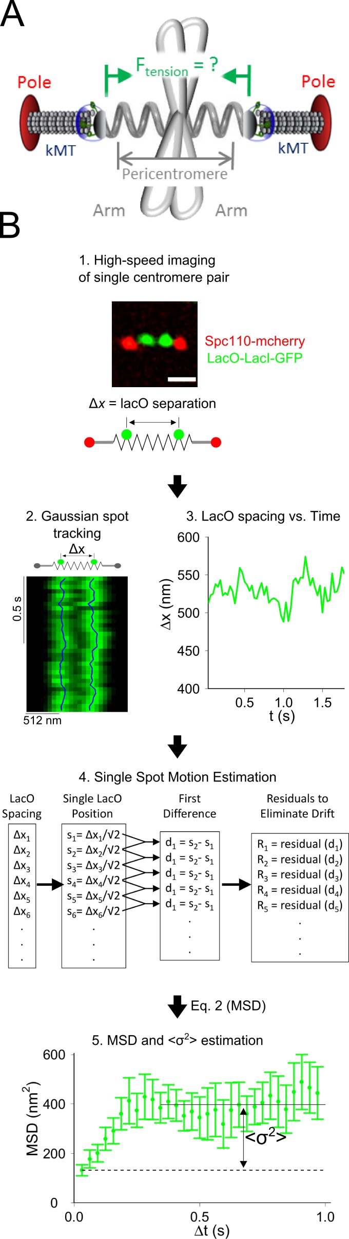
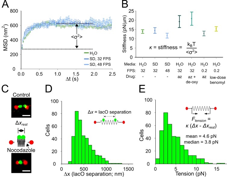
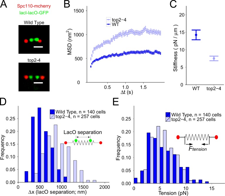
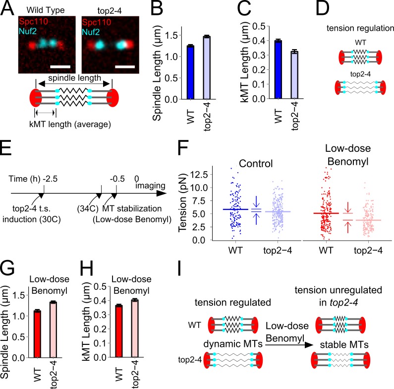
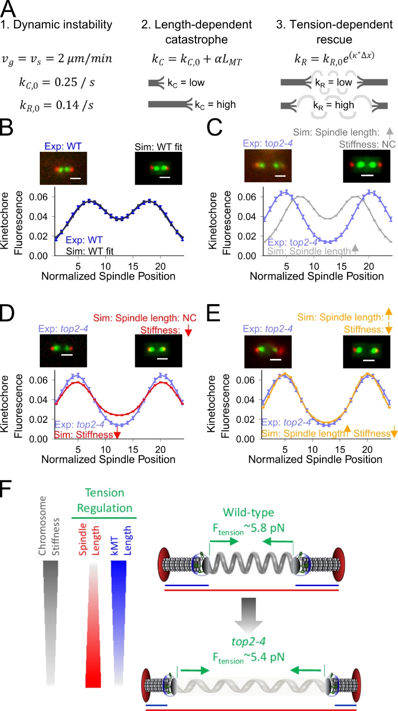
During cell division, a mitotic spindle is built by the cell and acts to align and stretch duplicated sister chromosomes before their ultimate segregation into daughter cells. Stretching of the pericentromeric chromatin during metaphase is thought to generate a tension-based signal that promotes proper chromosome segregation. However, it is not known whether the mitotic spindle actively maintains a set point tension magnitude for properly attached sister chromosomes to facilitate robust mechanochemical checkpoint signaling. By imaging and tracking the thermal movements of pericentromeric fluorescent markers in Saccharomyces cerevisiae, we measured pericentromere stiffness and then used the stiffness measurements to quantitatively evaluate the tension generated by pericentromere stretch during metaphase in wild-type cells and in mutants with disrupted chromosome structure. We found that pericentromere tension in yeast is substantial (4-6 pN) and is tightly self-regulated by the mitotic spindle: through adjustments in spindle structure, the cell maintains wild-type tension magnitudes even when pericentromere stiffness is disrupted.


Figures

Figure 1.
Figure 1.
In vivo pericentromere stiffness measurement. (A) Cartoon of yeast metaphase spindle denoting pericentromere location and tension. (B, 1) Gaussian-filtered experimental yeast metaphase spindle with fluorescent tags: LacO/lacI-GFP (lacO spots) and Spc110-mCherry (poles). Bar, 1 µm. (2) lacO spots were tracked using Gaussian mixture model fitting (blue lines). (3) The distance between the lacO spots (Δxi) over time. (4) Estimation of single spot motion and drift correction. (5) Eq. 2 converts Ri values to MSD, and then, the maximum motion (<σ2>) is estimated. Error bars = SEMs.
Figure 2.
Figure 2.
Substantial pericentromere stretching tension in budding yeast metaphase. (A) MSD versus time step size in WT cells (W303 strain background). (B) Mean pericentromere stiffness using different treatments. az = azide. (C) Estimating pericentromere rest length using nocodazole (see Materials and methods). Bars, 1 μm. (D) lacO spot separation distances in live cells (n = 390). (E) Pericentromere tension (n = 390). Error bars = SEMs.
Figure 3.
Figure 3.
Tension regulation in yeast metaphase spindles. (A) WT and top2-4 spindles, labeled as in Fig. 1. Bars, 1 μm. (B) MSD versus time step size in WT (light blue) and top2-4 (dark blue) cells. (C) Pericentromere stiffness. (D) lacO spot separation distances in WT cells and in top2-4 cells (P < 0.0001, t test). (E) Pericentromere tension distributions are similar between WT and top2-4 cells (P = 0.12, t test). Error bars = SEMs.
Figure 4.
Figure 4.
Tension is regulated by spindle structure. (A) Spindle and kMT lengths were measured in cells labeled with Spc110-mCherry and Nuf2-GFP. Bars, 1 μm. (B and C) Spindle length (B) and kMT length (C) in top2-4 and WT cells. (D) Tension regulation in top2-4 is accomplished via shorter kMT lengths and longer spindle lengths. (E) Experimental method to test whether dynamic kMTs are required to regulate tension in top2-4 cells. (F) In untreated cells, WT tension is maintained regardless of pericentromere stiffness (blue), but in cells treated with low-dose benomyl, which stabilizes kMTs, tension is reduced in top2-4 (red; control: n = 140 [WT] and n = 257 [top2-4]; benomyl: n = 215 [WT] and n = 197 [top2-4]). Horizontal lines and arrows are arithmetic means. (G) In benomyl, the relative difference in spindle length between WT and top2-4 is similar to untreated cells (vs. B). (H) In contrast, kMTs after benomyl treatment are longer in top2-4 (vs. C). (I) Tension regulation in top2-4 is disrupted when kMTs are stabilized in benomyl. Error bars = SEMs.
Figure 5.
Figure 5.
In simulations, tension-dependent kMT dynamics can explain tension regulation. (A) kMT dynamics in the simulation are governed by: (1) kMT dynamic instability with constant growth (vg) and shortening (vs) rates and with basal catastrophe (kC,0) and rescue (kR,0) frequencies; (2) kMT catastrophe frequency that increases with kMT length; and (3) kMT rescue frequency that increases with pericentromere tension. (B–E) Comparison of experimental images to simulated images convolved from the model (red, Spc110-mCherry; green, Nuf2-GFP). Bars, 1 µm. Exp = experimental image; Sim = simulated image; NC = no change. (B) WT experimental results compared with WT simulations. (C) top2-4 experimental results compared with simulations using longer top2-4 experimental spindle lengths with WT pericentromere stiffness. (D) top2-4 experimental results compared with simulations using reduced pericentromere stiffness and WT experimental spindle lengths. (E) top2-4 experimental results compared with simulations using reduced pericentromere stiffness and longer top2-4 experimental spindle lengths. (F) Pericentromere tension is regulated by spindle structure: increased stretch of the softer top2-4 pericentromere (light gray spring) relative to a stiffer WT pericentromere (dark gray) leads to similar tension (Ftension) because of increased spindle lengths and reduced kMT lengths. Error bars = SEMs.

References

    1. Akiyoshi B., Sarangapani K.K., Powers A.F., Nelson C.R., Reichow S.L., Arellano-Santoyo H., Gonen T., Ranish J.A., Asbury C.L., Biggins S. 2010. Tension directly stabilizes reconstituted kinetochore-microtubule attachments. Nature. 468:576–579 10.1038/nature09594 - DOI - PMC - PubMed
    1. Alexander S.P., Rieder C.L. 1991. Chromosome motion during attachment to the vertebrate spindle: initial saltatory-like behavior of chromosomes and quantitative analysis of force production by nascent kinetochore fibers. J. Cell Biol. 113:805–815 10.1083/jcb.113.4.805 - DOI - PMC - PubMed
    1. Asbury C.L., Gestaut D.R., Powers A.F., Franck A.D., Davis T.N. 2006. The Dam1 kinetochore complex harnesses microtubule dynamics to produce force and movement. Proc. Natl. Acad. Sci. USA. 103:9873–9878 10.1073/pnas.0602249103 - DOI - PMC - PubMed
    1. Bakhoum S.F., Thompson S.L., Manning A.L., Compton D.A. 2009. Genome stability is ensured by temporal control of kinetochore-microtubule dynamics. Nat. Cell Biol. 11:27–35 10.1038/ncb1809 - DOI - PMC - PubMed
    1. Bouck D.C., Bloom K. 2007. Pericentric chromatin is an elastic component of the mitotic spindle. Curr. Biol. 17:741–748 10.1016/j.cub.2007.03.033 - DOI - PMC - PubMed

Publication types

MeSH terms

Substances

LinkOut - more resources