. 2014 Jun 2;211(6):1049-62.
doi: 10.1084/jem.20131751. Epub 2014 May 12.

A novel Aβ-fibrinogen interaction inhibitor rescues altered thrombosis and cognitive decline in Alzheimer's disease mice

Affiliations

A novel Aβ-fibrinogen interaction inhibitor rescues altered thrombosis and cognitive decline in Alzheimer's disease mice

Hyung Jin Ahn et al. J Exp Med. .

Abstract

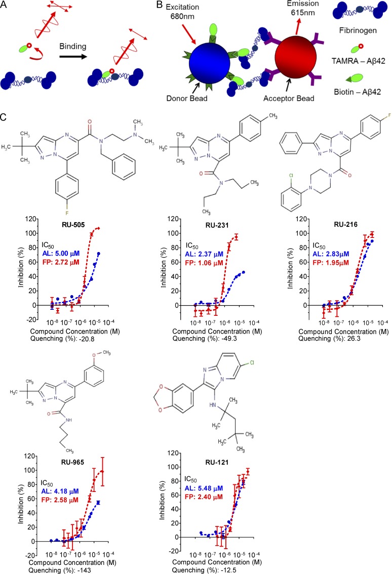
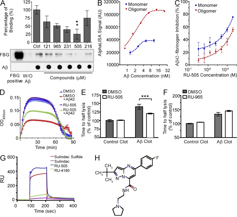
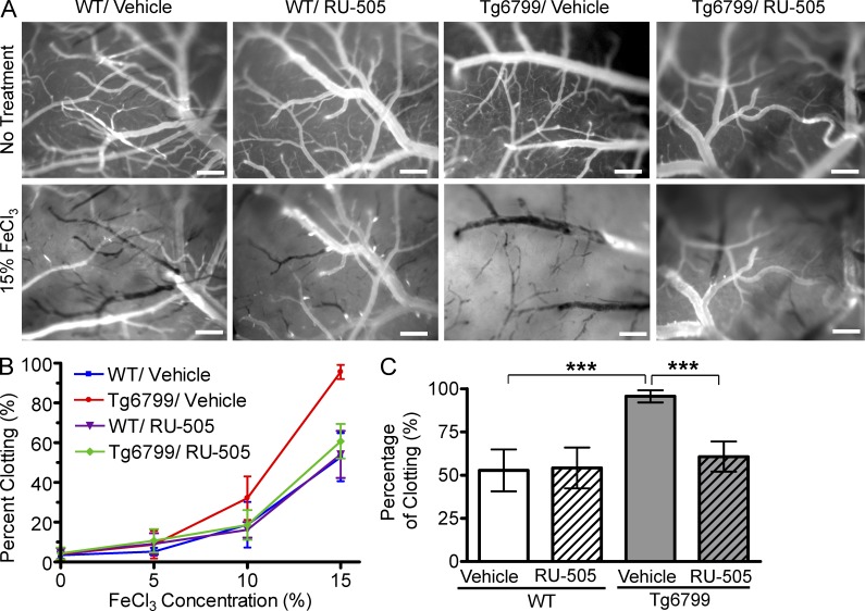
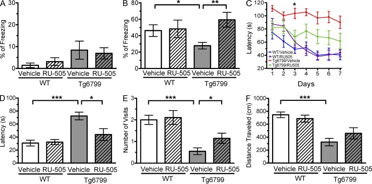
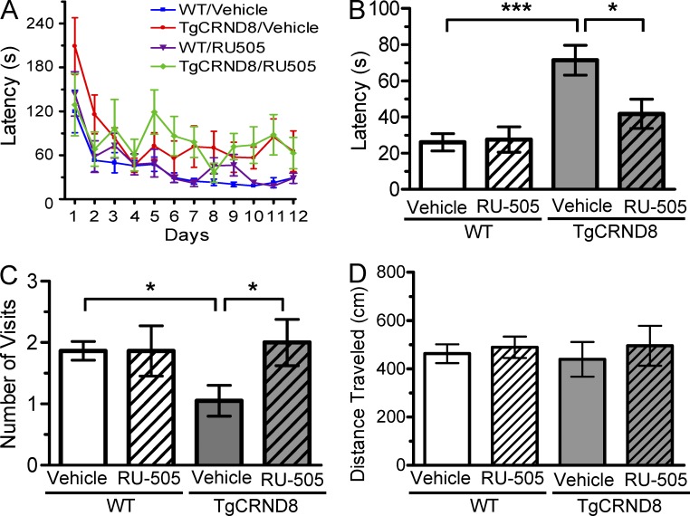
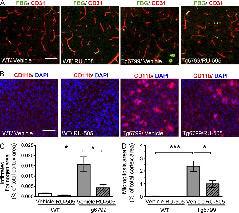
Many Alzheimer's disease (AD) patients suffer from cerebrovascular abnormalities such as altered cerebral blood flow and cerebral microinfarcts. Recently, fibrinogen has been identified as a strong cerebrovascular risk factor in AD, as it specifically binds to β-amyloid (Aβ), thereby altering fibrin clot structure and delaying clot degradation. To determine if the Aβ-fibrinogen interaction could be targeted as a potential new treatment for AD, we designed a high-throughput screen and identified RU-505 as an effective inhibitor of the Aβ-fibrinogen interaction. RU-505 restored Aβ-induced altered fibrin clot formation and degradation in vitro and inhibited vessel occlusion in AD transgenic mice. Furthermore, long-term treatment of RU-505 significantly reduced vascular amyloid deposition and microgliosis in the cortex and improved cognitive impairment in mouse models of AD. Our studies suggest that inhibitors targeting the Aβ-fibrinogen interaction show promise as therapy for treating AD.


Figures

Figure 1.
Figure 1.
The chemical structure and dose–response curve of Aβ–fibrinogen interaction inhibitors. (A) TAMRA–labeled Aβ peptide was bound to fibrinogen and the test compound, and the anisotropy of TAMRA–Aβ–fibrinogen binding was determined by FP. (B) Biotin-labeled Aβ42, which binds a streptavidin donor, was incubated with fibrinogen, which binds a protein A acceptor bead coated with antifibrinogen antibody. Aβ42 and fibrinogen interactions bring the beads in close proximity, resulting in the excitation of the donor beads and release of singlet oxygen molecules that triggers light emission in acceptor beads (AlphaLISA [AL]). (C) The half-maximal inhibitory concentration (IC50) values of the indicated compounds were determined by dose–response FP and AL experiments and are indicated inside the panel (red, FP; blue, AL). A quenching test was also performed to calculate how much each hit compound interfered with the AL signal at 10 µM concentration. Quenching values are indicated below the dose–response curve. n = 3–4 repeats per assay and all error bars indicate SEM. Data are representative of at least three independent experiments.
Figure 2.
Figure 2.
RU-505 inhibited the Aβ–fibrinogen interaction and restored Aβ-induced altered fibrin clot formation and degradation. (A) Candidate compounds (10 µM) were incubated with biotinylated Aβ42 and fibrinogen, and pull-down assays were performed using streptavidin–Sepharose. All samples were analyzed by Western blot. Dot blots were performed to control for amounts of Aβ pulled down. Control (Ctrl) lane contains only Aβ and fibrinogen without any compound (one-way ANOVA and Bonferroni post-hoc test; *, P < 0.05; n = 3–4 independent experiments). (B) The binding affinity between fibrinogen and monomeric or oligomeric biotinylated Aβ42 was measured using the AL assay. (n = 3–4 experiments, data are representative of three independent experiments). (C) The inhibitory efficacy of RU-505 on the interaction between fibrinogen and monomeric or oligomeric biotin-LC-Aβ42 was accessed in dose–response experiments using the AL assay. (n = 3–4 experiments, data are representative of three independent experiments). (D) RU-505 or DMSO was incubated with fibrinogen in the presence or absence of Aβ42, followed by plasminogen, thrombin, tPA, and CaCl2. Fibrin clot formation was assessed by measuring turbidity (n = 3 experiments, data are representative of three independent experiments). (E and F) The time to fibrin clot degradation was analyzed by measuring time to half lysis. Control clot half lysis time was set to 100% for each experiment and all other values were calculated relative to controls. (***, P < 0.001; n = 3 experiments, data are representative of three independent experiments). (G and H) Aβ42 was immobilized on the SPR sensor chip surface, and the interaction of the indicated compounds with Aβ42 was analyzed using Biacore 3000. Sulindac sulfide (known to bind Aβ42) was a positive control, and sulindac was negative control. (H) Chemical structure of RU-4180. Data are representative of three to four independent experiments. All values are means and SEM.
Figure 3.
Figure 3.
RU-505 prevented altered thrombosis and fibrinolysis in AD transgenic mice. (A) After craniotomy, three concentrations of FeCl3 (5, 10, and 15%) were incrementally administered to the surface of the brains of vehicle- or RU-505–treated WT and Tg6799 mice (Videos 1–4), and clotting of cerebral blood vessels (>20 µM) was imaged (bars, 200 µm). Representative intravital images shows the surface of the brains of vehicle- or RU-505–treated WT and Tg6799 mice before FeCl3 treatments or 5 min after 15% FeCl3 treatments. (B and C) Frequency of clotted vessels was calculated at increasing concentrations of FeCl3 (B) and was plotted for 15% FeCl3 treatment (C; ***, P < 0.001; n = 5 mice per group). All values are means and SEM. Results are from two independent experiments.
Figure 4.
Figure 4.
CAA pathology in AD transgenic mice was reduced after long-term treatment with RU-505. (A) Aβ deposits within cortical blood vessels of vehicle- or RU-505–treated Tg6799 mice were visualized using Congo red and laminin (green) labeling (bars, 100 µm). (B) Representative pictures showing parenchymal Aβ deposition in untreated and treated mice (bars, 100 µm). (C and D) CAA and Aβ plaques in (A) and (B) were quantified from 7–10 sections per mouse (n = 5 mice per group; *, P < 0.05). All values are means and SEM. Results are from two independent experiments.
Figure 5.
Figure 5.
RU-505 restored cognitive function in Tg6799 mice. (A) Freezing behavior was measured before electric foot shock during the training day to assess the basal freezing tendency of each group of mice. (n = 8–10 mice per group). (B) Contextual memory was assessed by measuring freezing behavior upon reexposure to the training chamber 24 h after fear conditioning training. (*, P < 0.05; **, P < 0.01; n = 8–10 mice per group). Results are from two independent experiments. (C–E) Spatial learning and memory retention of WT and Tg6799 mice was assessed using the Barnes maze after 3 mo of treatment with RU-505 or vehicle. One target hole was connected to a hidden escape chamber. (C) During training trials, latency to poke the target hole was measured. Significance was assessed using two-way ANOVA analysis with repeated measure (WT/vehicle vs. Tg6799/vehicle: F[1,120] = 40.47; P < 0.001; Tg6799/vehicle vs. Tg6799/RU-505: F[1,108] = 11.97; P < 0.01; n = 10–14 mice per group). Differences in latency were assessed by Bonferroni post hoc analysis. (D–F) During the Barnes maze probe trial, latency to reach the closed target hole (D), number of visits to the target hole (E), and total traveled distance (F) were measured ([E] *, P < 0.05; **, P < 0.01; ***, P < 0.001; n = 10–14 mice per group; [F] ***, P < 0.001; n = 10–14 mice per group). All results of the Barnes maze are from three independent experiments.
Figure 6.
Figure 6.
RU-505 restored spatial retention memory in TgCRND8 mice without affecting motor behavior. (A) The spatial memory of vehicle- or RU-505–treated WT and TgCRND8 mice was assessed using Barnes maze. (B and C) Spatial memory of RU-505–treated WT and TgCRND8 mice was tested using the Barnes maze probe trials. Time to reach the target hole (B), the number of visits to the closed target hole (C), and total distance traveled (D) were assessed (n = 7–11 mice per group). The results corroborate those in Fig. 5 and are from one experiment. All values are means and SEM.
Figure 7.
Figure 7.
Long-term treatment with RU-505 reduced the level of infiltrated fibrinogen and microgliosis in the cortex of Tg6799 mice. (A) Fibrinogen localized outside of endothelial cells of blood vessels was labeled with FITC-conjugated antifibrinogen antibody (green), and endothelial cells were labeled using anti-CD31 antibody (red; bars, 50 µm). (B) Activated microglia were visualized by staining for CD11b (red). DAPI staining (blue) was used to show integrity of tissue (bars, 100 µm). (C and D) Total fibrinogen area (C) and microgliosis (D) were quantified from 3 sections per mouse (n = 3–4 mice per group; *, P < 0.05; ***, P < 0.001). All values are means and SEM. Results are from two independent experiments.

Comment in

References

    1. Adams R.A., Bauer J., Flick M.J., Sikorski S.L., Nuriel T., Lassmann H., Degen J.L., Akassoglou K. 2007. The fibrin-derived gamma377-395 peptide inhibits microglia activation and suppresses relapsing paralysis in central nervous system autoimmune disease. J. Exp. Med. 204:571–582 10.1084/jem.20061931 - DOI - PMC - PubMed
    1. Ahn H.J., Zamolodchikov D., Cortes-Canteli M., Norris E.H., Glickman J.F., Strickland S. 2010. Alzheimer’s disease peptide beta-amyloid interacts with fibrinogen and induces its oligomerization. Proc. Natl. Acad. Sci. USA. 107:21812–21817 10.1073/pnas.1010373107 - DOI - PMC - PubMed
    1. Bayer T.A., Buslei R., Havas L., Falkai P. 1999. Evidence for activation of microglia in patients with psychiatric illnesses. Neurosci. Lett. 271:126–128 10.1016/S0304-3940(99)00545-5 - DOI - PubMed
    1. Brundel M., de Bresser J., van Dillen J.J., Kappelle L.J., Biessels G.J. 2012. Cerebral microinfarcts: a systematic review of neuropathological studies. J. Cereb. Blood Flow Metab. 32:425–436 10.1038/jcbfm.2011.200 - DOI - PMC - PubMed
    1. Chen Y.W., Gurol M.E., Rosand J., Viswanathan A., Rakich S.M., Groover T.R., Greenberg S.M., Smith E.E. 2006. Progression of white matter lesions and hemorrhages in cerebral amyloid angiopathy. Neurology. 67:83–87 10.1212/01.wnl.0000223613.57229.24 - DOI - PMC - PubMed

Publication types

MeSH terms