. 2014 Jun 15;192(12):5476-80.
doi: 10.4049/jimmunol.1400499. Epub 2014 May 12.

Cutting Edge: RIP1 kinase activity is dispensable for normal development but is a key regulator of inflammation in SHARPIN-deficient mice

Affiliations

Cutting Edge: RIP1 kinase activity is dispensable for normal development but is a key regulator of inflammation in SHARPIN-deficient mice

Scott B Berger et al. J Immunol. .

Abstract

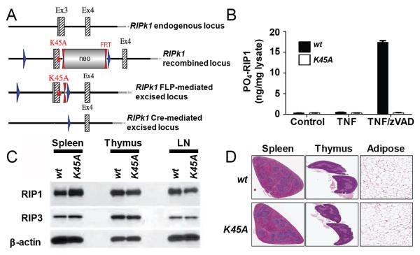
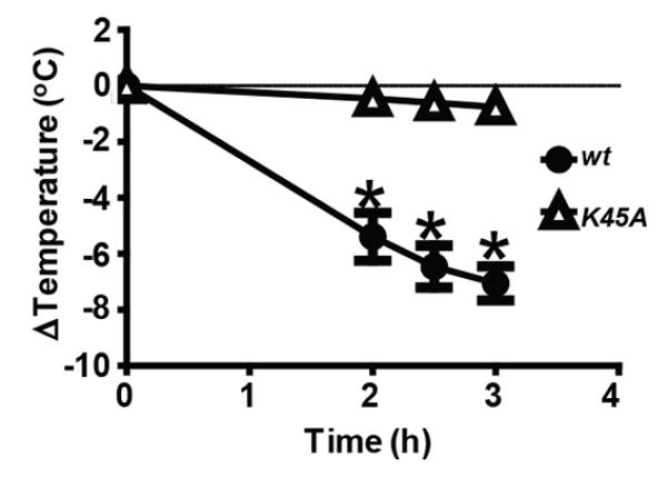
RIP1 (RIPK1) kinase is a key regulator of TNF-induced NF-κB activation, apoptosis, and necroptosis through its kinase and scaffolding activities. Dissecting the balance of RIP1 kinase activity and scaffolding function in vivo during development and TNF-dependent inflammation has been hampered by the perinatal lethality of RIP1-deficient mice. In this study, we generated RIP1 kinase-dead (Ripk1(K45A)) mice and showed they are viable and healthy, indicating that the kinase activity of RIP1, but not its scaffolding function, is dispensable for viability and homeostasis. After validating that the Ripk1(K45A) mice were specifically protected against necroptotic stimuli in vitro and in vivo, we crossed them with SHARPIN-deficient cpdm mice, which develop severe skin and multiorgan inflammation that has been hypothesized to be mediated by TNF-dependent apoptosis and/or necroptosis. Remarkably, crossing Ripk1(K45A) mice with the cpdm strain protected against all cpdm-related pathology. Together, these data suggest that RIP1 kinase represents an attractive therapeutic target for TNF-driven inflammatory diseases.


Figures

Figure 1
Figure 1
Ripk1K45A mice are viable and lack pathology associated with the RIP1 KO mouse. (A) The Ripk1 gene-targeting vector was constructed from genomic C57Bl/6 mouse DNA. The K45A point mutation was inserted into Ripk1 exon 3 while a Neo cassette was inserted in intron 3 flanked by FRT sites for Flp-mediated excision. Exon 3 was flanked by loxP sites enabling access to its deletion through Cre-action. (B) Phospho-RIP1 ELISA analysis. Lysates were prepared from wt (black bars) or Ripk1K45A (white bars) macrophages (control), or cells stimulated with TNF or TNF and zVAD for 3 hours. Results are representative of two experiments. (C) Western blot analyses of RIP1 and RIP3 expression in the spleen, thymus, and lymph nodes (LN) from wt and Ripk1K45A mice. β-Actin is shown as a loading control. Data are representative of three experiments. (D) Histopathology analysis of spleen, thymus and white adipose tissue from 10 to 12 week old wt littermate and Ripk1K45A mice. Data are representative of three wt and 5 Ripk1K45A mice.
Figure 2
Figure 2
Macrophages from Ripk1K45A mice are protected from necroptosis, but not NFκB activation or apoptosis in vitro. (A) CTG viability analysis of BMDM or TM from wt (black bars) or Ripk1K45A (white bars) mice. Macrophages were left untreated (control) or stimulated with TNF and zVAD. (B) Measurement of KC release from BMDM treated as in (A). (C) KC mRNA and protein analysis from BMDM from wt or Ripk1K45A mice treated with TNF and zVAD for the indicated times (D) CTG viability analysis of BMDM from wt (black bars) or Ripk1K45A (white bars) mice. Macrophages were either left untreated (control) or stimulated with TNF or TNF and CHX for 21 hours. (E) Measurement of KC release from BMDM treated as in (D).
Figure 3
Figure 3
Ripk1K45A mice are protected from TNF and zVAD-induced shock. wt (closed circles) or Ripk1K45A (open triangles) mice were injected with TNF and zVAD. Temperature was monitored over three hours, and animals were euthanized after a 7°C loss. Data are representative of at least 3 experiments, each containing seven mice per group.
Figure 4
Figure 4
RIP1 kinase activity drives inflammation in cpdm mice. (A) Representative photo of Ripk1K45A, cpdm and Ripk1K45A × cpdm mice at 7-8 weeks of age. (B) Cpdm and Ripk1K45A heterozygous mice were interbred to produce cpdm mice that were wt (WT/WT, black circles), heterozygous (WT/K45A, grey circles) or homozygous (K45A/K45A, white circles) for the Ripk1K45A gene. Dermatitis was considered to be severe (covering ≥ 50% of the abdomen). * indicates significantly different from RIP1 WT/WT (black circles) group. (C) Representative histology from cpdm or Ripk1K45A × cpdm mice at 7-8 weeks of age. Arrowheads in each panel point to, respectively, epidermal ulceration (black arrowhead) and dermal inflammation (red arrowhead) in skin, regions of inflammation in joint, regions of inflammation in lung and areas of inflammation in the liver. (D) Analysis of serum IgM levels.

References

    1. Carswell EA, Old LJ, Kassel RL, Green S, Fiore N, Williamson B. An endotoxin-induced serum factor that causes necrosis of tumors. Proc. Natl. Acad. Sci. U. S. A. 1975;72:3666–3670. - PMC - PubMed
    1. Aggarwal BB, Gupta SC, Kim JH. Historical perspectives on tumor necrosis factor and its superfamily: 25 years later, a golden journey. Blood. 2012;119:651–665. - PMC - PubMed
    1. Festjens N, Vanden Berghe T, Cornelis S, Vandenabeele P. RIP1, a kinase on the crossroads of a cell’s decision to live or die. Cell Death. Differ. 2007;14:400–410. - PubMed
    1. Vandenabeele P, Declercq W, Van HF, Vanden Berghe T. The role of the kinases RIP1 and RIP3 in TNF-induced necrosis. Sci. Signal. 2010;3:re4. - PubMed
    1. Degterev A, Hitomi J, Germscheid M, Ch’en IL, Korkina O, Teng X, Abbott D, Cuny GD, Yuan C, Wagner G, Hedrick SM, Gerber SA, Lugovskoy A, Yuan J. Identification of RIP1 kinase as a specific cellular target of necrostatins. Nat. Chem. Biol. 2008;4:313–321. - PMC - PubMed

Publication types

MeSH terms

Substances