

The pilot study of fibrin with temporomandibular joint derived synovial stem cells in repairing TMJ disc perforation
- PMID: 24822210
- PMCID: PMC4009306
- DOI: 10.1155/2014/454021
The pilot study of fibrin with temporomandibular joint derived synovial stem cells in repairing TMJ disc perforation
Abstract
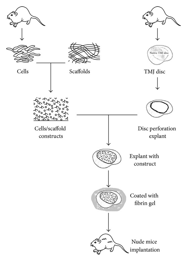
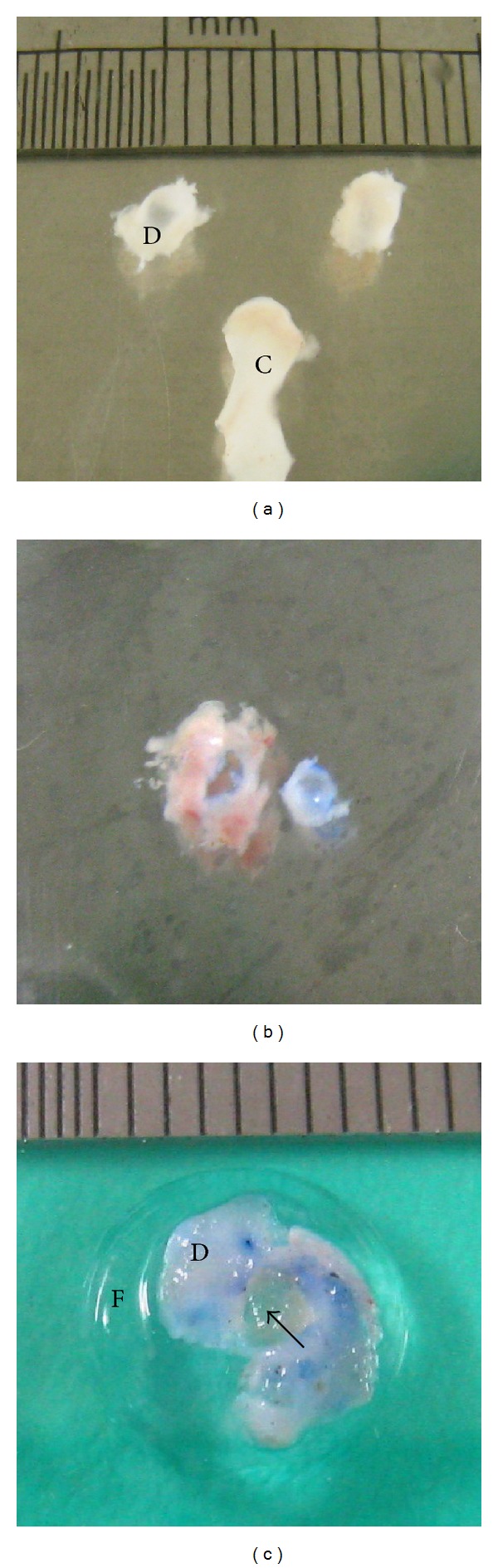
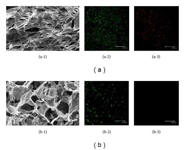
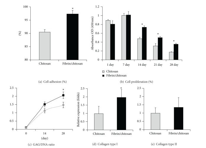
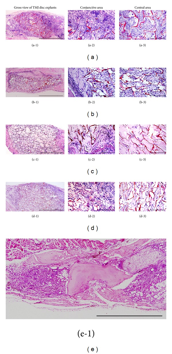
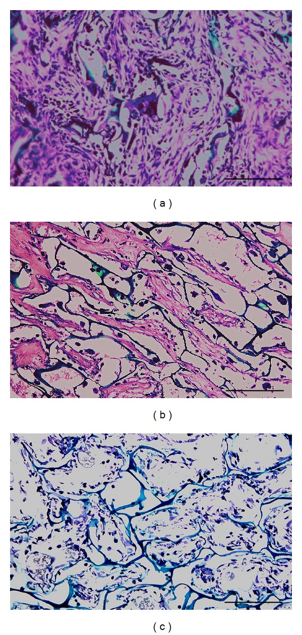
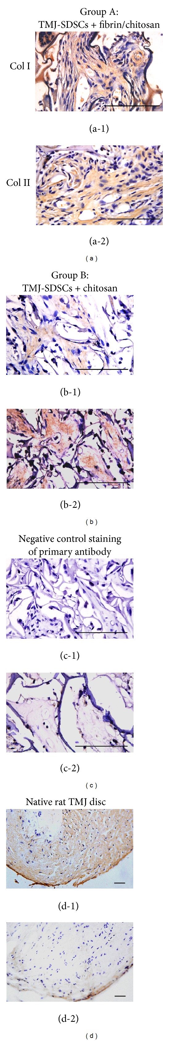
TMJ disc related diseases are difficult to be cured due to the poor repair ability of the disc. TMJ-SDSCs were ideal cell sources for cartilage tissue engineering which have been widely used in hyaline cartilage regeneration. Fibrin gel has been demonstrated as a potential scaffold for neocartilage formation. The aim of this study was to repair the TMJ disc perforation using fibrin/chitosan hybrid scaffold combined with TMJ-SDSCs. Rat TMJ-SDSCs were cultured on hybrid scaffold or pure chitosan scaffolds. The cell seeding efficiency, distribution, proliferation, and chondrogenic differentiation capacity were investigated. To evaluate the in vivo repair ability of cell/scaffold construct, rat TMJ disc explants were punched with a defect to mimic TMJ disc perforation. Cell seeded scaffolds were inserted into the defect of TMJ disc explants and then were implanted subcutaneously in nude mice for 4 weeks. Results demonstrated that fibrin may improve cell seeding, proliferation, and chondrogenic induction in vitro. The in vivo experiments showed more cartilage ECM deposition in fibrin/chitosan scaffold, which suggested an enhanced reparative ability. This pilot study demonstrated that the regenerative ability of TMJ-SDSCs seeded in fibrin/chitosan scaffold could be applied for repairing TMJ disc perforation.
Figures
Similar articles
-
Fibro/chondrogenic differentiation of dental stem cells into chitosan/alginate scaffolds towards temporomandibular joint disc regeneration.J Mater Sci Mater Med. 2018 Jun 26;29(7):97. doi: 10.1007/s10856-018-6109-6. J Mater Sci Mater Med. 2018. PMID: 29946796
-
An asymmetric chitosan scaffold for tendon tissue engineering: In vitro and in vivo evaluation with rat tendon stem/progenitor cells.Acta Biomater. 2018 Jun;73:377-387. doi: 10.1016/j.actbio.2018.04.027. Epub 2018 Apr 17. Acta Biomater. 2018. PMID: 29678676
-
[In vitro and in vivo study of chondrogenesis on the hybrid scaffold from fibrin modified PLGA and adipose-derived stem cells].Xi Bao Yu Fen Zi Mian Yi Xue Za Zhi. 2010 Aug;26(8):758-60. Xi Bao Yu Fen Zi Mian Yi Xue Za Zhi. 2010. PMID: 21032949 Chinese.
-
Specific tissue engineering for temporomandibular joint disc perforation.Cytotherapy. 2024 Mar;26(3):231-241. doi: 10.1016/j.jcyt.2023.11.005. Epub 2023 Dec 14. Cytotherapy. 2024. PMID: 38099894 Review.
-
Temporomandibular Joint Regenerative Medicine.Int J Mol Sci. 2018 Feb 2;19(2):446. doi: 10.3390/ijms19020446. Int J Mol Sci. 2018. PMID: 29393880 Free PMC article. Review.
Cited by
-
Mesenchymal Stromal Cell Transplantation Induces Regeneration of Large and Full-Thickness Cartilage Defect of the Temporomandibular Joint.Cartilage. 2021 Dec;13(1_suppl):1814S-1821S. doi: 10.1177/1947603520926711. Epub 2020 Jun 4. Cartilage. 2021. PMID: 32493042 Free PMC article.
-
Stem Cells for Temporomandibular Joint Repair and Regeneration.Stem Cell Rev Rep. 2015 Oct;11(5):728-42. doi: 10.1007/s12015-015-9604-x. Stem Cell Rev Rep. 2015. PMID: 26123357 Review.
-
Biomimetic Scaffolds for Regeneration of Temporomandibular Joint Disc: A Narrative Review.J Dent (Shiraz). 2024 Jun 1;25(2):108-117. doi: 10.30476/dentjods.2023.97625.2024. eCollection 2024 Jun. J Dent (Shiraz). 2024. PMID: 38962074 Free PMC article. Review.
-
Mesenchymal stem cells-derived microvesicles versus platelet-rich plasma in the treatment of monoiodoacetate-induced temporomandibular joint osteoarthritis in Albino rats.Heliyon. 2022 Oct 1;8(10):e10857. doi: 10.1016/j.heliyon.2022.e10857. eCollection 2022 Oct. Heliyon. 2022. PMID: 36212013 Free PMC article.
-
Research progress on tissue engineering in repairing tempomandibular joint.Zhejiang Da Xue Xue Bao Yi Xue Ban. 2021 Apr 25;50(2):212-221. doi: 10.3724/zdxbyxb-2021-0118. Zhejiang Da Xue Xue Bao Yi Xue Ban. 2021. PMID: 34137227 Free PMC article. English.
References
-
- Jibiki M, Shimoda S, Nakagawa Y, Kawasaki K, Asada K, Ishibashi K. Calcifications of the disc of the temporomandibular joint. Journal of Oral Pathology and Medicine. 1999;28(9):413–419. - PubMed
-
- Mercuri LG, Wolford LM, Sanders B, Dean White R, Giobbie-Hurder A. Long-term follow-up of the CAD/CAM patient fitted total temporomandibular joint reconstruction system. Journal of Oral and Maxillofacial Surgery. 2002;60(12):1440–1448. - PubMed
-
- Allen KD, Athanasiou KA. Scaffold and growth factor selection in temporomandibular joint disc engineering. Journal of Dental Research. 2008;87(2):180–185. - PubMed
-
- Allen KD, Athanasiou KA. Growth factor effects on passaged TMJ disk cells in monolayer and pellet cultures. Orthodontics & Craniofacial Research. 2006;9(3):143–152. - PubMed
MeSH terms
Substances
LinkOut - more resources
Full Text Sources
Other Literature Sources
Medical
