Review
. 2014;11(6):673-81.
doi: 10.4161/rna.28985. Epub 2014 May 7.

Versatile microRNA biogenesis in animals and their viruses

Affiliations
Review

Versatile microRNA biogenesis in animals and their viruses

Mingyi Xie et al. RNA Biol. 2014.

Abstract

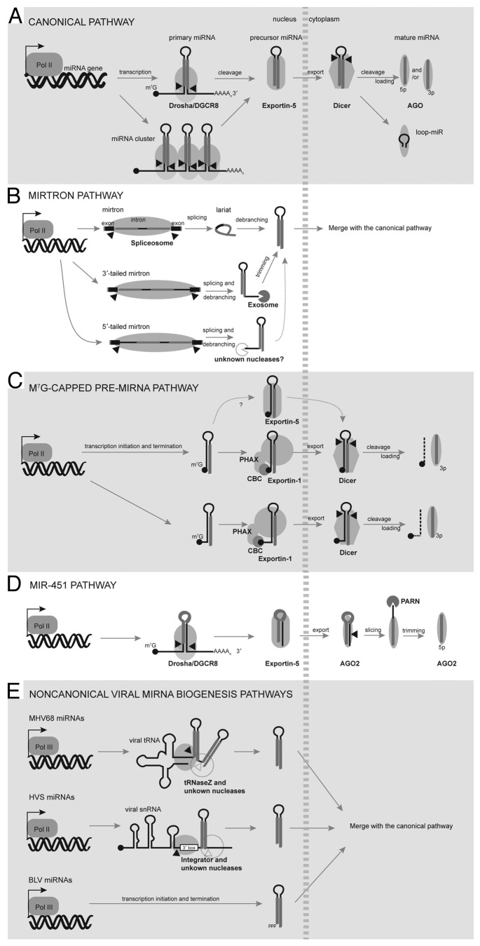
MicroRNAs (miRNAs) are ubiquitous gene regulators that modulate essential cellular processes at the post-transcriptional level. In metazoans and their viruses, most miRNAs are produced from hairpin-containing primary transcripts that are sequentially cleaved by nuclear Drosha and cytoplasmic Dicer. In the last decade, alternative mechanisms that bypass either the Drosha or Dicer cleavage step have emerged, increasing the complexity of the miRNA regulatory network. Here, we highlight non-canonical pathways that generate miRNAs using a variety of molecular machineries that play fundamental roles in the biogenesis and processing of other classes of cellular RNAs.

Keywords: AGO2 slicer activity; Dicer; Integrator; Microprocessor; exportin-5; m7G-capped pre-miRNA; microRNA biogenesis; mirtron; tRNaseZ; viral microRNA.


Figures

None
Figure 1. MiRNA biogenesis pathways in animals and their viruses. (A) The canonical pathway. Pri-miRNAs containing a single or a cluster of hairpins are typically transcribed by Pol II, 5′-capped, and 3′-polyadenylated. The Microprocessor complex (Drosha and DGCR8) cleaves pri-miRNAs to release the pre-miRNA hairpins, which are exported by XPO5 and processed by Dicer into mature miRNA duplexes in the cytoplasm. One miRNA strand is preferably selected by AGO to form RISC. In certain cases, the loop of the pre-miRNA hairpin may be incorporated into RISC. (B) The mirtron pathway. Microprocessor-independent mirtrons are directly generated by splicing of short introns and the resulting lariats are debranched to form pre-miRNA hairpins. Mirtrons could have tails on either end of the pre-miRNA hairpins. 3′-tailed mirtrons are further trimmed by the exosome, while the nuclease that processes 5′-tailed mirtrons remains elusive. (C) The m7G-capped pre-miRNA pathway. M7G-capped pre-miRNAs are directly transcribed by Pol II, bypassing Microprocessor cleavage. The presence of the 5′ cap directs pre-miRNAs to the PHAX-XPO1 export pathway, known to function in the export of snRNAs. However, except for the 5′-tailed variants, the m7G-capped pre-miRNAs retain the ability to be exported by XPO5. After Dicer cleavage, the 5′-capped 5p-miRNA is unable to associate effectively with AGO, resulting in the production of only 3p-miRNPs. (D) The miR-451 pathway. Drosha cleavage of pri-miR-451 releases an unusual pre-miRNA that is too short to be processed by Dicer. Instead, AGO2 cleaves the 3p-arm of pre-miR-451 and PARN trims to generate a 5p-miRNA. (E) Drosha-independent miRNA biogenesis in animal viruses. MHV68 pri-miRNAs are tRNA-pre-miRNA chimeras that are processed by tRNaseZ at the 5′ end of the first pre-miRNA hairpin. The enzyme that separates the two pre-miRNAs is unknown. HVS pri-miRNAs are snRNA-pre-miRNA chimeras that are processed by Integrator to release the pre-miRNA. The 3′-end formation mechanism for HVS pre-miRNAs remains elusive. BLV miRNAs are derived from pre-miRNAs that are directly transcribed by Pol III as endogenous shRNAs. All viral pre-miRNAs described here are exported by XPO5 and processed by Dicer.
None
Figure 2. Mouse pre-miR-484 is a 5′-tailed m7G-capped pre-miRNA. (A) The histogram shows cap-seq reads mapping to the mmu-miR-484 locus. TSS and transcription directionality are indicated by a black arrow. The height of each bar is proportional to the normalized read number (raw read number per million mapped reads). Cap-seq and miRNA-seq data were taken from refs. 9 and 66, respectively. (B) Mouse pre-miR-484 is illustrated with miR-484 highlighted in gray. (C) Northern blot probed for miR-484 of 10 µg total RNA isolated from mouse astrocyte DBT cells. M, in vitro-transcribed m7G-capped pre-miR-484 markers with or without a 3-nt AAG 5′-end extension. (D) Xenopus oocyte microinjection assay. A mixture of 1–10 fmoles 32P-labeled EBER-1, U1∆Sm, tRNAᵖʰᵉ, and pre-miR-15b with or without 1 pmole of unlabeled pre-miR-484 (m7G-capped and containing 5′-AAG) was injected into the nuclei of X. laevis oocytes. After 2-h incubation, six oocytes were manually dissected, and RNAs from the nucleus (N) and cytoplasm (C) equivalent to one oocyte were extracted and analyzed on an 8M urea-15% polyacrylamide gel. inj, injected material. The bar graph shows relative RNA export efficiency (cytoplasmic/total) of the indicated RNAs with pre-miR-484 competition (black bars) compared with no competition (white bars). Error bars represent standard deviation from two experiments. (E) In vitro-transcribed pre-miR-484 with or without a 3-nt AAG 5′-end extension was incubated with recombinant human Dicer (a generous gift from Dr Jennifer Doudna) for various times and analyzed by northern blot to detect miR-484 (top panel) or miR-484-5p (lower panel), using 32P-labeled DNA oligos complementary to miR-484 or miR-484-5p, respectively. The blot shows cleavage of pre-miR-484 without the 5′-end extension (☐) or pre-miR-484 (x) by purified human Dicer at each time point. Error bars represent standard deviations in two experiments. The Materials and Methods used here were essentially the same as described in reference .

References

    1. Bartel DP. MicroRNAs: target recognition and regulatory functions. Cell. 2009;136:215–33. doi: 10.1016/j.cell.2009.01.002. - DOI - PMC - PubMed
    1. Krol J, Loedige I, Filipowicz W. The widespread regulation of microRNA biogenesis, function and decay. Nat Rev Genet. 2010;11:597–610. - PubMed
    1. Huntzinger E, Izaurralde E. Gene silencing by microRNAs: contributions of translational repression and mRNA decay. Nat Rev Genet. 2011;12:99–110. doi: 10.1038/nrg2936. - DOI - PubMed
    1. Fabian MR, Sonenberg N. The mechanics of miRNA-mediated gene silencing: a look under the hood of miRISC. Nat Struct Mol Biol. 2012;19:586–93. doi: 10.1038/nsmb.2296. - DOI - PubMed
    1. Kozomara A, Griffiths-Jones S. miRBase: integrating microRNA annotation and deep-sequencing data. Nucleic Acids Res. 2011;39:D152–7. doi: 10.1093/nar/gkq1027. - DOI - PMC - PubMed

Publication types

LinkOut - more resources