. 2014 May 13;12(5):e1001860.
doi: 10.1371/journal.pbio.1001860. eCollection 2014 May.

RNA mimicry by the fap7 adenylate kinase in ribosome biogenesis

Affiliations

RNA mimicry by the fap7 adenylate kinase in ribosome biogenesis

Jérôme Loc'h et al. PLoS Biol. .

Abstract

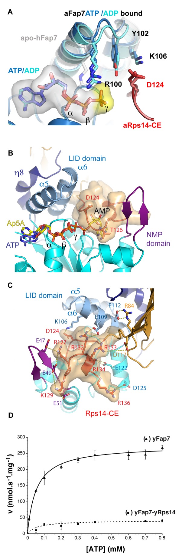
During biogenesis of the 40S and 60S ribosomal subunits, the pre-40S particles are exported to the cytoplasm prior to final cleavage of the 20S pre-rRNA to mature 18S rRNA. Amongst the factors involved in this maturation step, Fap7 is unusual, as it both interacts with ribosomal protein Rps14 and harbors adenylate kinase activity, a function not usually associated with ribonucleoprotein assembly. Human hFap7 also regulates Cajal body assembly and cell cycle progression via the p53-MDM2 pathway. This work presents the functional and structural characterization of the Fap7-Rps14 complex. We report that Fap7 association blocks the RNA binding surface of Rps14 and, conversely, Rps14 binding inhibits adenylate kinase activity of Fap7. In addition, the affinity of Fap7 for Rps14 is higher with bound ADP, whereas ATP hydrolysis dissociates the complex. These results suggest that Fap7 chaperones Rps14 assembly into pre-40S particles via RNA mimicry in an ATP-dependent manner. Incorporation of Rps14 by Fap7 leads to a structural rearrangement of the platform domain necessary for the pre-rRNA to acquire a cleavage competent conformation.


Conflict of interest statement

The authors have declared that no competing interests exist.

Figures

Figure 1
Figure 1. Structure of the aFap7–aRps14 complex.
(A) Cartoon representation of the aFap7–aRps14 complex in complex with ADP. aFap7 is represented in blue/purple shades and Rps14 in orange for the CORE domain and red for the C-terminal extension. (B) Superposition of the ADP (light blue) and ATP (dark blue) aFap7 structure with the hFap7 in complex with ADP-Pi (magenta, PDB 3iil). (C) Superposition of aRps14 in complex with aFap7 (orange of the core domain, red for the Rps14-CE, and brown for the β4–α2 loop), with yRps14 in complex with the ribosome (green)
Figure 2
Figure 2. SAXS structure of the yeast and archaeal complexes.
SAXS scattering profiles of (A) the aFap7–aRps14 complexes and (B) yeast yFap7–yRps14 complexes and (C) free yFap7. (Left) The experimental scattering profile is depicted in dashed red lines and the calculated profile from the best fit in blue lines. The residual is depicted below the scattering curve. The initial scattering profile of the initial yFap7 model is represented in green. (Right) Representation of the calculated envelope with the best fit model. Rps14 is represented in yellow and Fap7 in blue, except in (C) where yFap7 is colored blue (N terminus) to red (C terminus).
Figure 3
Figure 3. Both the Rps14 and Fap7 C-ter domain participate in their interaction.
To analyze Fap7 and Rps14 interaction and the domains involved, their association was observed by analytical gel filtration. Note that the C-terminal deletion of aFap7 removes the only tryptophane, which leads to the reduced absorption in the gel-filtration profile. (A) Gel filtration profiles of aRps14 (black), aFap7 (dotted line), and a 1∶1 mix of aRps14–aFap7 (blue). (B) Gel filtration profiles of aRps14 (black), aFap7ΔC (dotted line), and a 1∶1 mix of aFap7ΔC–aRps14 (green). (C) Gel filtration profiles of aRps14ΔC (black), aFap7 (dotted line), and a 1∶1 mix of aFap7–aRps14ΔC (red). (D) Gel filtration profiles of aRps14ΔC (black), aFap7ΔC (dotted line), and a 1∶1 mix of aFap7ΔC–aRps14ΔC (blue).
Figure 4
Figure 4. Rps14 obstructs the Fap7 AMP binding cavity.
(A) Conformational differences of the ATP binding pocket in complex with ADP (light blue) and ATP (dark blue). The apo-hFap7 structure is represented in grey. (B) Superposition of Ap5A with ATP from AK1 (PDB code 1z83) in the aFap7–aRps14 complex defines the AMP binding pocket. This pocket is filled by the aRps14 C-terminal extension. (C) Cartoon representation of the aFap7–aRps14 complex in the putative AMP substrate binding cavity of aFap7. The C-terminal extension of Rps14 is represented with a transparent surface. Salt bridges to basic residues are indicated. (D) Comparison of AK activities with respect to ATP at a constant concentration of AMP (0.3 mM) of yFap7 (▴) and of the yRps14–yFap7 complex (▪).
Figure 5
Figure 5. yFap7 acts as an RNA mimic.
(A) yRps14 in the yeast ribosome . The 40S is colored green and 60S blue. The Rps26 and Rps1 proteins have been omitted for clarity. Helix 23, 34, and 45 are colored light green, dark green, and pink, respectively. (B) Close-up view of the yRps14 interaction with RNA. The Fap7 protein placed in the same position as in the aFap7–aRps14 complex is overlaid in blue surface representation (middle and right). (C) Secondary structure of helix 23 from S. cerevisiae 18S rRNA (nucleotides 884 to 928 with GGG extension at 5′ end) used for binding assays. (D) Quantification of bound RNA ratio versus total RNA in each well and nonlinear curve fitting of the data points. Interaction of RNA with yRps14 (•), yRps14–yFap7 complex (▪), and yFap7 (▴). (E) Primer extension analysis showing the activation of in vitro site-D cleavage in pre-40S particles purified, using PTH-NOB1 from either wild-type cells (WT) or after depletion of Fap7 (PGAL::FAP7), by nucleotide addition. The strong upper stops result from termination at the sites of 18S rRNA base-methylation at A1779 and A1780. These modifications precede site-D cleavage in vivo. The arrow indicates site D. ATP was added at 1 mM when indicated. We added 250 pmoles of recombinant Fap7-his or Fap7–Rps14 complex when indicated.
Figure 6
Figure 6. Fap7 ATPase activity regulates its association with Rsp14.
Interaction between yFap7 and GST-Rps14 was tested by pulldown experiments. (A) Interaction of yFap7 with GST-Rps14 was tested by addition of 800 pmoles of yFap7 on GST-yRps14 beads resuspended in 1 ml of IP buffer. Effect of addition of ATP, ADP, or AMP-PNP at 1 mM final concentration in the presence of MgCl2 (5 mM) was tested. Protein associated with the beads was analyzed by Coomassie staining. For input controls, 10% of Fap7 (80 pmoles) and the same quantity of Rps14 beads used for the IP were loaded. (B) Effects of magnesium and nucleotide concentration were tested by using the same strategy in the presence of RNA (cf., D). Two quantities of nucleotides were used: 1 µmole (1 mM) or 10 nmole (10 µM). Experiments were done in the presence or in the absence of 5 mM MgCl2. (C) Association of GST-yRps14 to RNA was assessed by a competition experiment using a different ratio between RNA and yFap7: 800 pmole Fap7 with 800 pmole RNA (1∶1), 400 pmole Fap7 with 800 pmole RNA (1∶2), and 200 pmole Fap7 with 800 pmole RNA (1∶4). Nucleotides were added at 1 mM final. Protein associated with the beads was analyzed by Coomassie staining. For input controls, 10% of Fap7 (80 pmoles) and the same quantity of Rps14 beads used for the IP were loaded. (D) Same as in C, but this time the RNA counterpart was followed on Urea-acrylamide gel after SYBR Safe staining. For input control, 80 pmole of RNA was loaded. (E) ATPase activity of yFap7 (black points) was followed by a coupled enzyme assay. Effect of addition after 10 min of MBP-yRps14 (black line), buffer (dotted dark line), or Prp43 (dotted dark grey line) was also monitored. In parallel, ATPase activity of MBP-yRps14 (light grey line) alone and the preformed complex yFap7–yRps14 (dark grey line) was also tested.
Figure 7
Figure 7. Model of Fap7 function in ribosome biogenesis.
(A) Model of the conformational change leading to Rps14 release. The two conformations of the aFap7 NMP domain, the Rps14-CE and Rps14 β4-α2 loop, are represented in red and green. The remaining aFap7 is in surface representation. The seven disordered C-terminal residues of the green conformation are depicted by a dashed line. The extrapolated movement of the three regions suggested to accompany disruption of the Fap7–Rps14 complex is indicated by black arrows (see also Movie S1). (B) During ribosome maturation, Fap7 and Rps14 are bound in the platform domain, which is in a nonnative conformation. After A2 cleavage, Fap7 removes Rps14 from the rRNA. Rps14 is then dissociated from Fap7, probably following ATP hydrolysis, and reincorporated in a near cognate position. This enables a major conformational change to occur that restructures the platform domain and the ITS1, delivering a pre-40S that can undergo the final cleavage at site D. In the absence of Fap7, Rps14 cannot be reincorporated in the ribosome, the rRNA in the platform domain is not remodeled, and cleavage of the D site is not possible, leading to a dead end in the pathway.

Similar articles

Cited by

References

    1. Kressler D, Hurt E, Bassler J (2010) Driving ribosome assembly. Biochim Biophys Acta BBA - Mol Cell Res 1803: 673–683. - PubMed
    1. Panse VG, Johnson AW (2010) Maturation of eukaryotic ribosomes: acquisition of functionality. Trends Biochem Sci 35(5): 260–266. - PMC - PubMed
    1. Karbstein K (2011) Inside the 40S ribosome assembly machinery. Curr Opin Chem Biol 15: 657–663. - PMC - PubMed
    1. Lebaron S, Schneider C, van Nues RW, Swiatkowska A, Walsh D, et al. (2012) Proofreading of pre-40S ribosome maturation by a translation initiation factor and 60S subunits. Nat Struct Mol Biol 19: 744–753. - PMC - PubMed
    1. Strunk BS, Novak MN, Young CL, Karbstein K (2012) Aac translation-like cycle is a quality control checkpoint for maturing 40S ribosome subunits. Cell 150: 111–121. - PMC - PubMed

Publication types

MeSH terms