. 2014 Jul;55(7):1298-309.
doi: 10.1194/jlr.M048207. Epub 2014 May 13.

Ceramide kinase is required for a normal eicosanoid response and the subsequent orderly migration of fibroblasts

Affiliations

Ceramide kinase is required for a normal eicosanoid response and the subsequent orderly migration of fibroblasts

Dayanjan S Wijesinghe et al. J Lipid Res. 2014 Jul.

Abstract

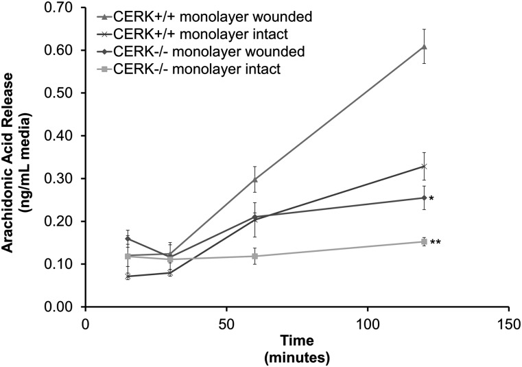
In these studies, the role of ceramide-1-phosphate (C1P) in the wound-healing process was investigated. Specifically, fibroblasts isolated from mice with the known anabolic enzyme for C1P, ceramide kinase (CERK), ablated (CERK(-/-) mice) and their wild-type littermates (CERK(+/+)) were subjected to in vitro wound-healing assays. Simulation of mechanical trauma of a wound by scratching a monolayer of fibroblasts from CERK(+/+) mice demonstrated steadily increasing levels of arachidonic acid in a time-dependent manner in stark contrast to CERK(-/-) fibroblasts. This observed difference was reflected in scratch-induced eicosanoid levels. Similar, but somewhat less intense, changes were observed in a more complex system utilizing skin biopsies obtained from CERK-null mice. Importantly, C1P levels increased during the early stages of human wound healing correlating with the transition from the inflammatory stage to the peak of the fibroplasia stage (e.g., proliferation and migration of fibroblasts). Finally, the loss of proper eicosanoid response translated into an abnormal migration pattern for the fibroblasts isolated from CERK(-/-) As the proper migration of fibroblasts is one of the necessary steps of wound healing, these studies demonstrate a novel requirement for the CERK-derived C1P in the proper healing response of wounds.

Keywords: ceramide-1-phosphate; lipidomics; wound healing.


Figures

Fig. 1.
Fig. 1.
Genetic ablation of CERK translates to a significant loss of specific forms of C1P. Immortalized embryonic fibroblasts (2 × 10⁶) from both CERK+/+ and CERK−/− backgrounds were seeded onto 100 mm tissue culture plates and allowed to grow to confluency overnight under standard incubation conditions. Lipids were extracted from these cells, and the lipids species of interest were quantified via HPLC ESI-MS/MS as described in Experimental Procedures. C1P levels are represented in units of picomoles per 1 × 10⁶ cells and are the means of n = 6 of individual experiments ± SD. A one-way ANOVA followed by a Tukey’s post hoc test was used to assess statistical significance (** P < 0.01).
Fig. 2.
Fig. 2.
Genetic ablation of CERK and the resultant loss of CERK-derived C1P adversely affects AA release by fibroblasts in response to mechanical trauma. Immortalized embryonic fibroblasts (2 × 10⁶) from both CERK+/+ and CERK−/− backgrounds were seeded onto 100 mm tissue culture plates and allowed to grow to confluency overnight under standard incubation conditions in medium containing [3H] AA as described in Experimental Procedures. Following the removal of unlabeled AA, the cells were first rested for 2 h in medium containing 2% serum, and the monolayers were either left unharmed as controls or subjected to scratch-induced mechanical trauma as described in Experimental Procedures. The medium was collected at the indicated time points and counted via scintillation. The data are presented as nanograms of AA released per milliliter of medium and are the average of three different experiments ± SD. A one-way ANOVA followed by a Tukey’s post hoc test was used to assess statistical significance of the AA released by the different groups. The analysis revealed significant differences in AA release between wounded CERK+/+ and CERK−/− fibroblasts (* P < 0.05, ** P < 0.01).
Fig. 3.
Fig. 3.
Genetic ablation of CERK and the resultant loss of CERK-derived C1P adversely affects eicosanoid synthesis by fibroblasts in response to mechanical trauma. A–H: Immortalized embryonic fibroblasts (2 × 10⁶) from both CERK+/+ and CERK−/− backgrounds were seeded onto 100 mm tissue culture plates and allowed to grow to confluency overnight under standard incubation conditions. Following overnight incubation, the cells were rested for 2 h in medium containing 2% serum, and the monolayers were either left unharmed as controls or subjected to scratch-induced mechanical trauma as described in Experimental Procedures. The eicosanoids secreted into the medium in response to scratch-induced mechanical trauma were quantified at 2 h and 4 h postinjury via HPLC ESI-MS/MS as described in Experimental Procedures. A–H: Quantification of AA, PGE2, PGF2α, 6-keto PGF1α, 5-HETE, 11-HETE, 12-HETE, and 15-HETE, respectively, expressed in units of nanograms lipid per milliliter medium. The data presented are the average of three individual experiments performed on two separate occasions ± SD. Student’s t-test was used to assess statistical significance of the difference in scratch-induced eicosanoid release between CERK+/+ and CERK−/− fibroblasts (* P < 0.05, ** P < 0.01).
Fig. 4.
Fig. 4.
CERK-derived C1P is required for eicosanoid release in response to wounding of the skin. Punch biopsies (10 mm) were obtained from the shaved and depilated dorsum of CERK+/+ and CERK −/− mice according to Experimental Procedures, followed by incubation for 2 h and 4 h under standard incubator conditions in medium containing 2% serum as described in Experimental Procedures. The eicosanoids secreted into the medium from the injured skin were quantified at 2 h and 4 h postbiopsy via HPLC ESI-MS/MS as described in Experimental Procedures. A–H: Quantification of AA, PGE2, 6-keto PGF1α, PGF2α, 5-HETE, 11-HETE, 12-HETE, and 15-HETE, respectively, expressed in units of nanograms lipid per square centimeter excised skin. The data presented are the average of three individual experiments performed on two separate occasions ± SD. Student’s t-test was used to assess statistical significance of eicosanoid synthesis by CERK+/+ skin biopsy compared with that of CERK−/− (* P < 0.05, ** P < 0.01).
Fig. 5.
Fig. 5.
Loss of CERK-derived C1P alters actin distribution and migration of fibroblasts. A–B: CERK+/+ and CERK−/− MEFs (0.2 × 10⁵ cells) were seeded overnight onto 22 × 22 mm coverslips in 35 mm plates and stained for actin and DNA as described in Experimental Procedures. C–F: Cells were seeded into 35 mm cell culture dishes and allowed to grow to confluency. Migration was initiated by scratching confluent cells with a p10 pipette tip as described in Experimental Procedures, and the motility was tracked as described in Experimental Procedures. C: Tracings of individual cell migration paths from immortalized CERK+/+ and CERK−/− following scratch-induced mechanical trauma. D: Tracings of individual cell migration paths from primary CERK+/+ and CERK−/− following scratch-induced mechanical trauma. E: Percent wound closure with time (upper panel) and average velocity of movement and meandering index (lower panels) of immortalized CERK+/+ and CERK−/− MEFs. F: Percent wound closure with time (upper panel) and average velocity of movement and meandering index (lower panels) of primary CERK+/+ and CERK−/− MEFs.
Fig. 6.
Fig. 6.
Abnormal migration of fibroblasts due to loss of CERK is partially rescued via the addition of exogenous eicosanoids. A: MEFs (2 × 10⁶) from both CERK+/+ and CERK−/− backgrounds were seeded onto 100 mm tissue culture plates and allowed to grow to confluency overnight under standard incubator conditions. Following overnight incubation, the cells were rested for 2 h in medium containing 2% serum. The monolayer was then subjected to scratch-induced mechanical trauma with a 200 μl pipette tip, and the medium was supplemented either with ethanol (vehicle) or the relevant deficient eicosanoids as described in Experimental Procedures. The migration of the fibroblasts to the cleared area was photographically recorded at 3 h postscratch. B-E: Cells were seeded into 35 mm cell culture dishes and allowed to grow to confluency and were rested for 2 h in medium containing 2% serum. The monolayer was then subjected to scratch-induced mechanical trauma cells with a p10 pipette tip, and the medium was supplemented either with ethanol (vehicle) or the relevant deficient eicosanoids as described in Experimental Procedures. Cell motility was tracked as described in Experimental Procedures using video microscopy. B: Percent wound closure with time. C: Average velocity of movement. D: Meandering index. E: Tracings of individual cell migration paths. The data presented are the average of six individual experiments performed on two separate occasions ± SD. A one-way ANOVA followed by a Tukey’s post hoc test was used to assess statistical significance of the migration between CERK+/+ and CERK−/− treatment groups (* P < 0.05, ** P < 0.01).
Fig. 7.
Fig. 7.
The levels of C1P increase during the early stages of wound healing in human patients. PTFE tubings (3 cm) expanded to 90 μm pore size were implanted subcutaneously in the upper arm using a 14 gauge needle as described in Experimental Procedures, thereby creating an acute wound. A punch biopsy (10 mm) was taken at the same time for comparison (day 0). The tubes were removed on days 3, 5, 7, and 14 allowing cells involved in wound healing to infiltrate. The lipids were extracted from 1 cm length of PTFE tubing and subjected to quantitation via HPLC ESI-MS/MS as described in Experimental Procedures. The data presented are the average from three individuals ± SD. Student’s t-test was used to assess statistical significance between C1P levels compared with day 0 (* P < 0.05).

Similar articles

Cited by

References

    1. Goldberg S. R., Diegelmann R. F. 2010. Wound healing primer. Surg. Clin. North Am. 90: 1133–1146 - PubMed
    1. Berry D. P., Harding K. G., Stanton M. R., Jasani B., Ehrlich H. P. 1998. Human wound contraction: collagen organization, fibroblasts, and myofibroblasts. Plast. Reconstr. Surg. 102: 124–131, discussion 132–134. - PubMed
    1. Xu F., Zhang C., Graves D. T. 2013. Abnormal cell responses and role of TNF-α in impaired diabetic wound healing. Biomed. Res. Int . 2013: 754802. - PMC - PubMed
    1. Suganuma H., Sato A., Tamura R., Chida K. 1995. Enhanced migration of fibroblasts derived from lungs with fibrotic lesions. Thorax. 50: 984–989 - PMC - PubMed
    1. White E. S., Atrasz R. G., Dickie E. G., Aronoff D. M., Stambolic V., Mak T. W., Moore B. B., Peters-Golden M. 2005. Prostaglandin E2 inhibits fibroblast migration by E-prostanoid 2 receptor–mediated increase in PTEN activity. Am. J. Respir. Cell Mol. Biol. 32: 135–141 - PMC - PubMed

Publication types

LinkOut - more resources