Review
. 2014 Sep 15;23(R1):R17-26.
doi: 10.1093/hmg/ddu204. Epub 2014 May 13.

Human-induced pluripotent stem cells: potential for neurodegenerative diseases

Affiliations
Review

Human-induced pluripotent stem cells: potential for neurodegenerative diseases

Christopher A Ross et al. Hum Mol Genet. .

Abstract

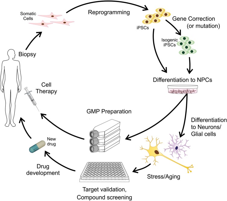
The cell biology of human neurodegenerative diseases has been difficult to study till recently. The development of human induced pluripotent stem cell (iPSC) models has greatly enhanced our ability to model disease in human cells. Methods have recently been improved, including increasing reprogramming efficiency, introducing non-viral and non-integrating methods of cell reprogramming, and using novel gene editing techniques for generating genetically corrected lines from patient-derived iPSCs, or for generating mutations in control cell lines. In this review, we highlight accomplishments made using iPSC models to study neurodegenerative disorders such as Huntington's disease, Parkinson's disease, Amyotrophic Lateral Sclerosis, Fronto-Temporal Dementia, Alzheimer's disease, Spinomuscular Atrophy and other polyglutamine diseases. We review disease-related phenotypes shown in patient-derived iPSCs differentiated to relevant neural subtypes, often with stressors or cell "aging", to enhance disease-specific phenotypes. We also discuss prospects for the future of using of iPSC models of neurodegenerative disorders, including screening and testing of therapeutic compounds, and possibly of cell transplantation in regenerative medicine. The new iPSC models have the potential to greatly enhance our understanding of pathogenesis and to facilitate the development of novel therapeutics.


Figures

Figure 1.
Figure 1.
Schematic diagram showing uses of human somatic cells such as adult skin fibroblasts reprogrammed into pluripotent stem cells (iPSCs). The iPSCs derived from a donor carrying a mutation in a disease risk gene can be genetically corrected using homologous recombination, ZFN, TALEN or CRISPR/Cas9. The resulting isogenic cells can be used as normal controls or potentially for cell transplantation therapy. These techniques can also be used to introduce disease-related mutations into the genomes of healthy donor derived iPSCs. The genetically modified cells and parental iPSCs can be differentiated into neural stem (precursor) cells (NSCs). The NSCs can be expanded, sorted for appropriate neural precursors, and further differentiated into mature disease-specific neural subtypes. For research into neural degenerative disorders, cells stressors or artificial cell aging may be applied to enhance disease-specific phenotypes. The cells can be used for compound screening for drug discovery and testing of novel therapeutics.
Figure 2.
Figure 2.
iPSCs derived from HD, PD and ALS patients have been successfully differentiated into mature neurons with relevant properties, and show disease-specific phenotypes. (AD). HD iPSC model (26). (A) HD iPSCs with 109 CAG repeats can be differentiated into mature, striatal-like neurons that express medium-spiny-neuron markers MAP2a/b and Bcl11B. (B) Time-lapse experiment. The risk of death was significantly higher for the HD180i.7 line (180 CAG) compared to the HD33i.8 line (33 CAG) over time in culture. After BDNF removal, the risk of death was significantly greater for the HD180i.7 line compared to the HD33i.8 line. (C) Hierarchical clustering of genes from striatal-like cells is represented by the vertical bars (yellow for HD and green for control). HD and control cells are clearly separated. (D) Quantification of condensed nuclei as a measure of cell toxicity showed that both HD109i.1 and HD180i.5 lines had significantly more cell death after BDNF withdrawal, whereas the HD33i.8 control line showed no change. (E–H). PD iPSC model (57). (E) Differentiation of iPSCs into midbrain dopaminergic (mDA) neurons, and expression of LRRK2 protein in dopaminergic neurons. (F–G) Electrophysiological recordings of control HUF5 and PD G2019S iPSCs differentiated to DA neurons demonstrates that they can fire action potential. (H) Quantification of cells double positive for TH and CASP3 in clones of normal (wild-type) H9 and HUF5-iPSC versus disease G2019S-iPSC-derived DA neurons, with several H2O2 concentrations. (IM). ALS-FTD model (75). (I) C9ORF72 ALS cells show cytoplasmic RAN translation peptides. C9ORF72 iPSN cells contain cytoplasmic GGGGCC RNA foci, while control iPSNs do not. (J) Quantification of cytoplasmic RNA foci reveals that cytoplasmic foci are RNase A sensitive. (K) C9ORF72 ALS iPSC neurons are highly susceptible to glutamate toxicity. Immunofluorescent staining of control and C9ORF72 iPSC cultures show expression of glutamate receptors GluR2, NR2B, and postsynaptic density protein PSD95 at comparable levels. Box indicates region of high magnification seen below each image. (L) Dose response curve of control and C9ORF72 iPSC neurons revealed that C9ORF72 iPSC neurons are highly susceptible to glutamate excitotoxicity. (M) Glutamate-induced excitotoxicity of C9ORF72 iPSNs shows statistically significant cell death after 4 hr of 30 mM glutamate treatment when compared to control iPSNs.

References

    1. Winner B., Marchetto M.C., Winkler J., Gage F.H. Human-induced pluripotent stem cells pave the road for a better understanding of motor neuron disease. Hum. Mol. Genet. 2014;23:R27–R34. - PubMed
    1. Jakel R.J., Schneider B.L., Svendsen C.N. Using human neural stem cells to model neurological disease. Nat. Rev. Genet. 2004;5:136–144. - PubMed
    1. Hargus G., Ehrlich M., Hallmann A.L., Kuhlmann T. Human stem cell models of neurodegeneration: a novel approach to study mechanisms of disease development. Acta Neuropathol. 2014;127:151–173. - PubMed
    1. Gage F.H., Temple S. Neural stem cells: generating and regenerating the brain. Neuron. 2013;80:588–601. - PubMed
    1. Qiang L., Fujita R., Abeliovich A. Remodeling neurodegeneration: somatic cell reprogramming-based models of adult neurological disorders. Neuron. 2013;78:957–969. - PubMed

Publication types