

Human heat shock protein-specific cytotoxic T lymphocytes display potent antitumour immunity in multiple myeloma
- PMID: 24824351
- PMCID: PMC4134698
- DOI: 10.1111/bjh.12943
Human heat shock protein-specific cytotoxic T lymphocytes display potent antitumour immunity in multiple myeloma
Abstract
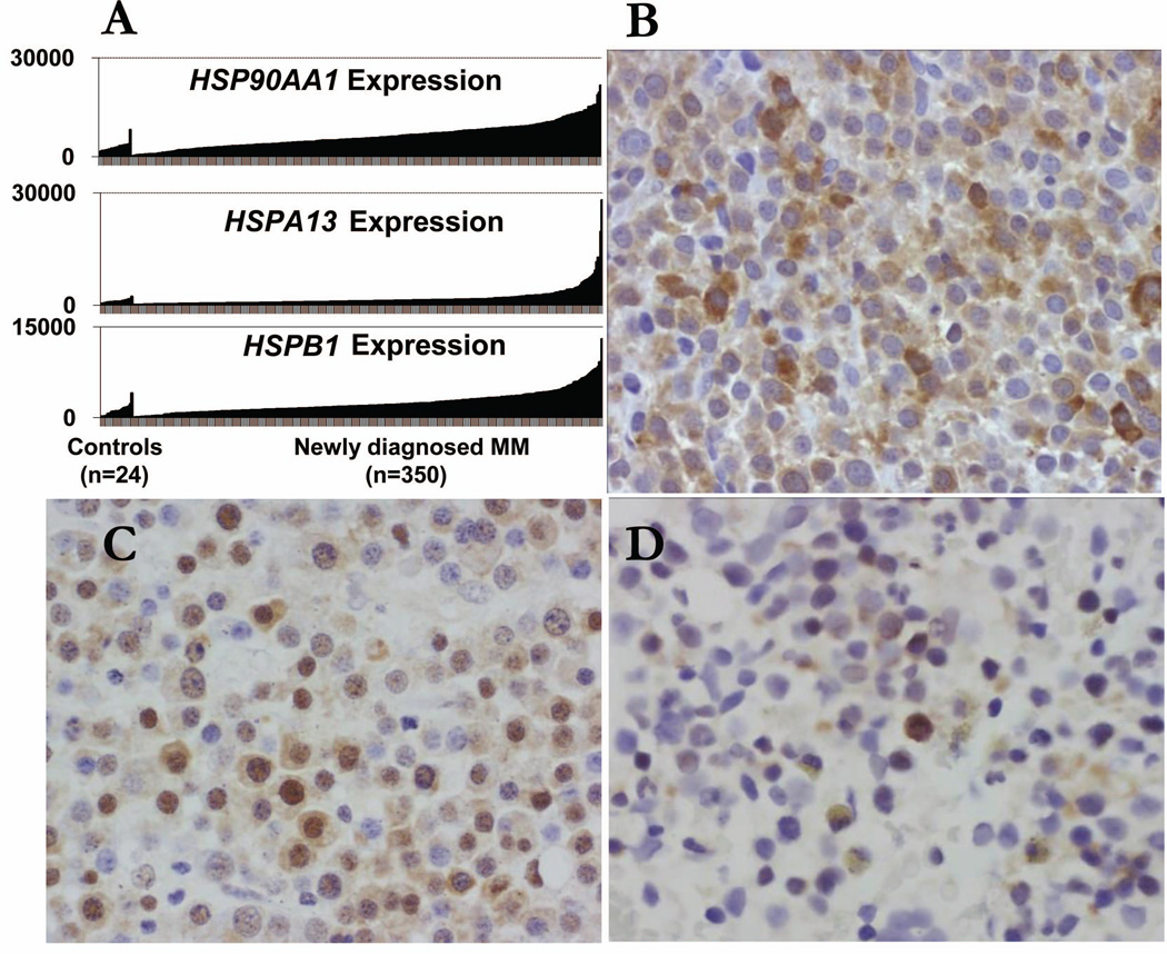
Tumour cell-derived heat shock proteins (HSPs) are used as vaccines for immunotherapy of cancer patients. However, it is proposed that the peptide chaperoned on HSPs, not HSPs themselves, elicited a potent immune response. Given that HSPs are highly expressed by most myeloma cells and vital to myeloma cell survival, we reasoned that HSPs themselves might be an ideal myeloma antigen. In the present study, we explored the feasibility of targeting HSPs themselves for treating multiple myeloma. We identified and chose HLA-A*0201-binding peptides from human HSPB1 (HSP27) and HSP90AA1 (HSP90), and confirmed their immunogenicity in HLA-A*0201 transgenic mice. Dendritic cells pulsed with HSPB1 and HSP90AA1 peptides were used to stimulate peripheral blood mononuclear cells from healthy volunteers and myeloma patients to generate HSP peptide-specific cytotoxic T lymphocytes (CTLs). HSP peptide-specific CTLs efficiently lysed HLA-A*0201(+) myeloma cells (established cell lines and primary plasma cells) but not HLA-A*0201(-) myeloma cells in vitro, indicating that myeloma cells naturally express HSP peptides in the context of major histocompatibility complex class I molecules. More importantly, HSP peptide-specific CTLs effectively reduced tumour burden in the xenograft mouse model of myeloma. Our study clearly demonstrated that HSPs might be novel tumour antigens for immunotherapy of myeloma.
Keywords: heat shock proteins; immunotherapy; multiple myeloma; vaccine.
© 2014 John Wiley & Sons Ltd.
Conflict of interest statement
Figures


Similar articles
-
Targeting heat shock proteins for immunotherapy in multiple myeloma: generation of myeloma-specific CTLs using dendritic cells pulsed with tumor-derived gp96.Clin Cancer Res. 2005 Dec 15;11(24 Pt 1):8808-15. doi: 10.1158/1078-0432.CCR-05-1553. Clin Cancer Res. 2005. PMID: 16361569
-
Identification of human leucocyte antigen (HLA)-A*0201-restricted cytotoxic T lymphocyte epitopes derived from HLA-DOβ as a novel target for multiple myeloma.Br J Haematol. 2013 Nov;163(3):343-51. doi: 10.1111/bjh.12544. Epub 2013 Aug 30. Br J Haematol. 2013. PMID: 24032635 Free PMC article.
-
Myeloma-specific multiple peptides able to generate cytotoxic T lymphocytes: a potential therapeutic application in multiple myeloma and other plasma cell disorders.Clin Cancer Res. 2012 Sep 1;18(17):4850-60. doi: 10.1158/1078-0432.CCR-11-2776. Epub 2012 Jul 2. Clin Cancer Res. 2012. PMID: 22753586 Free PMC article.
-
Antitumor cytotoxic T-lymphocyte response in human lung carcinoma: identification of a tumor-associated antigen.Immunol Rev. 2002 Oct;188:114-21. doi: 10.1034/j.1600-065x.2002.18810.x. Immunol Rev. 2002. PMID: 12445285 Review.
-
Immunotherapy for human cancer using heat shock protein-peptide complexes.Curr Oncol Rep. 2005 Mar;7(2):104-8. doi: 10.1007/s11912-005-0035-8. Curr Oncol Rep. 2005. PMID: 15717943 Review.
Cited by
-
Ex vivo Optimization of Glucose-Regulated Protein 94/Glycoprotein 96 Expressions in Mammospheres; Implication for Breast Cancer Immunotherapy.Cell J. 2022 May;24(5):261-266. doi: 10.22074/cellj.2022.7908. Epub 2022 Apr 27. Cell J. 2022. PMID: 35717566 Free PMC article.
-
Network pharmacology- and molecular docking-based investigation of the therapeutic potential and mechanism of daucosterol against multiple myeloma.Transl Cancer Res. 2023 Apr 28;12(4):1006-1020. doi: 10.21037/tcr-23-456. Transl Cancer Res. 2023. PMID: 37180669 Free PMC article.
-
Crossroad between the Heat Shock Protein and Inflammation Pathway in Acquiring Drug Resistance: A Possible Target for Future Cancer Therapeutics.Biomedicines. 2023 Sep 26;11(10):2639. doi: 10.3390/biomedicines11102639. Biomedicines. 2023. PMID: 37893013 Free PMC article. Review.
-
Unveiling the potential of HSPA4: a comprehensive pan-cancer analysis of HSPA4 in diagnosis, prognosis, and immunotherapy.Aging (Albany NY). 2024 Jan 31;16(3):2517-2541. doi: 10.18632/aging.205496. Epub 2024 Jan 31. Aging (Albany NY). 2024. PMID: 38305786 Free PMC article.
-
Phosphorylation of HSF1 at serine 326 residue is related to the maintenance of gynecologic cancer stem cells through expression of HSP27.Oncotarget. 2017 May 9;8(19):31540-31553. doi: 10.18632/oncotarget.16361. Oncotarget. 2017. PMID: 28415561 Free PMC article.
References
-
- Albrecht J, Frey M, Teschner D, Carbol A, Theobald M, Herr W, Distler E. IL-21-treated naive CD45RA+ CD8+ T cells represent a reliable source for producing leukemia-reactive cytotoxic T lymphocytes with high proliferative potential and early differentiation phenotype. Cancer Immunology Immunotherapy. 2011;60:235–248. - PMC - PubMed
-
- Alexander J, Oseroff C, Dahlberg C, Qin M, Ishioka G, Beebe M, Fikes J, Newman M, Chesnut RW, Morton PA, Fok K, Appella E, Sette A. A decaepitope polypeptide primes for multiple CD8+ IFN-gamma and Th lymphocyte responses: evaluation of multiepitope polypeptides as a mode for vaccine delivery. Journal of Immunology. 2002;168:6189–6198. - PubMed
Publication types
MeSH terms
Substances
Grants and funding
LinkOut - more resources
Full Text Sources
Other Literature Sources
Medical
Molecular Biology Databases
Research Materials
Miscellaneous
