

Bi-directional x-ray phase-contrast mammography
- PMID: 24824594
- PMCID: PMC4019485
- DOI: 10.1371/journal.pone.0093502
Bi-directional x-ray phase-contrast mammography
Abstract
Phase-contrast x-ray imaging is a promising improvement of conventional absorption-based mammography for early tumor detection. This potential has been demonstrated recently, utilizing structured gratings to obtain differential phase and dark-field scattering images. However, the inherently anisotropic imaging sensitivity of the proposed mono-directional approach yields only insufficient diagnostic information, and has low diagnostic sensitivity to highly oriented structures. To overcome these limitations, we present a two-directional x-ray phase-contrast mammography approach and demonstrate its advantages by applying it to a freshly dissected, cancerous mastectomy breast specimen. We illustrate that the two-directional scanning procedure overcomes the insufficient diagnostic value of a single scan, and reliably detects tumor structures, independently from their orientation within the breast. Our results indicate the indispensable diagnostic necessity and benefit of a multi-directional approach for x-ray phase-contrast mammography.
Conflict of interest statement
Figures

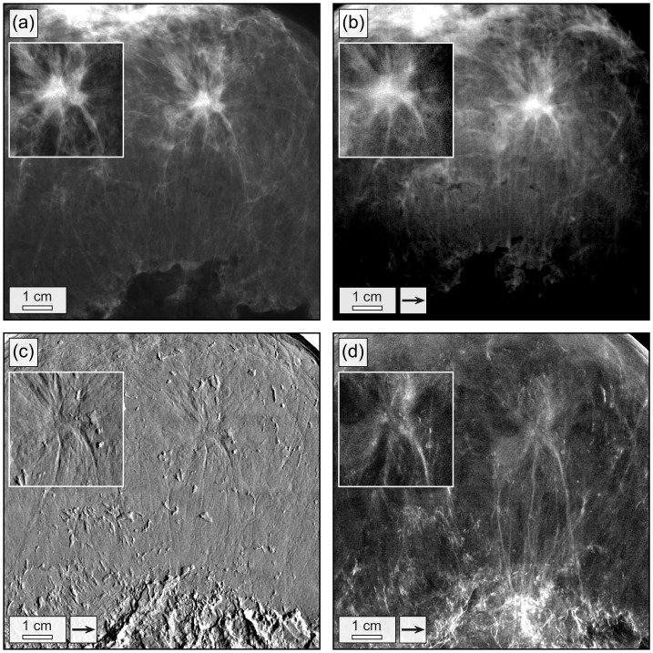
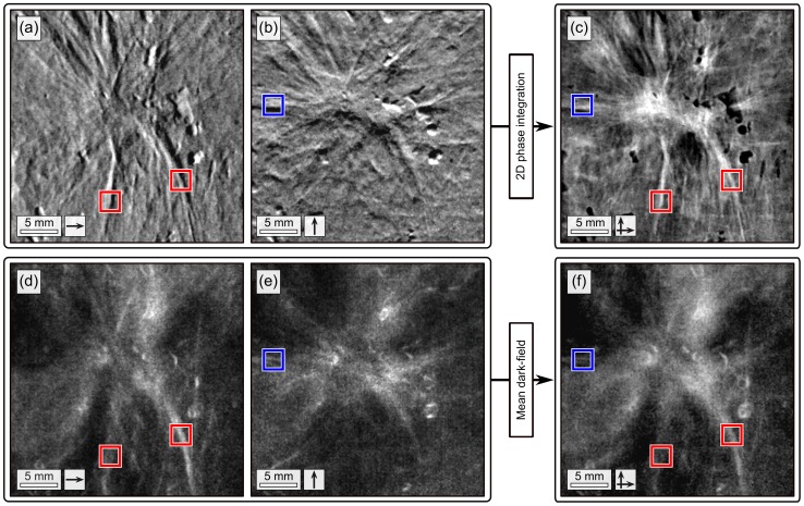
 (a),
(a),  (b) and sharpened, two-dimensional integrated phase image
  (c). Dark-field
  (d),
(d),  (e) and mean dark-field
  image (f). Arrows indicate the direction of scanning. Red and blue boxes indicate tumor branches exclusively perceivable in the images obtained with scanning performed in
  - or
- or  -direction, respectively.
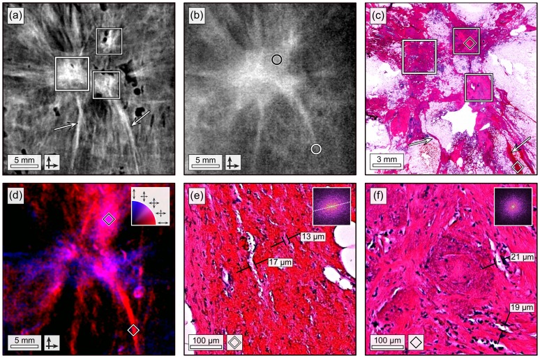
 of the invasive ductal carcinoma. The frames in (a) and (c) indicate three locally separated tumor lesions (apparently trifocal). Arrows in (a) and (c) indicate fine tumor branches. (b) Sharpened, absorption image
  of the invasive ductal carcinoma. Circles indicate position of relevant tumor details. (c) Histological slice (Hematoxylin and Eosin stain) of the invasive ductal carcinoma. (d) Directional dark-field image of the invasive ductal carcinoma. Preferred scattering direction is color-coded ranging from
  -directed (red) over isotropic (purple) to
  -directed (blue). (e) 200x magnified histological image of the tumor branch, as indicated by white diamond in (c) and (d). The 2-dimensional FFT is shown as an inlay. (d) 200x magnified histological image of the tumor lesion, as indicated by black diamond in (c) and (d). The 2-dimensional FFT is shown as an inlay.
References
-
- Siegel R, Naishadham D, Jemal A (2012) Cancer statistics, 2012. CA: A Cancer Journal for Clinicians 62: 1029. - PubMed
-
- Poplack SP, Tosteson AN, Grove MR, Wells WA, Carney PA (2000) Mammography in 53,803 women from the new hampshire mammography network. Radiology 217: 832–840. - PubMed
-
- Keyriläinen J, Bravin A, Fernandez M, Tenhunen M, Virkkunen P, et al. (2010) Phase-contrast x-ray imaging of breast. Acta radiologica 51: 866–884. - PubMed
-
- Takeda T, Momose A, Ueno E, Itai Y (1998) Phase-contrast x-ray CT image of breast tumor. Journal of Synchrotron Radiation 5: 1133–1135. - PubMed
-
- Arfelli F, Bonvicini V, Bravin A, Cantatore G, Castelli E, et al. (2000) Mammography with synchrotron radiation: phase-detection techniques. Radiology 215: 286–293. - PubMed
Publication types
MeSH terms
LinkOut - more resources
Full Text Sources
Other Literature Sources
Medical
