. 2014 May 13;5(3):e00935-14.
doi: 10.1128/mBio.00935-14.

Coordinating bacterial cell division with nutrient availability: a role for glycolysis

Affiliations

Coordinating bacterial cell division with nutrient availability: a role for glycolysis

Leigh G Monahan et al. mBio. .

Abstract

Cell division in bacteria is driven by a cytoskeletal ring structure, the Z ring, composed of polymers of the tubulin-like protein FtsZ. Z-ring formation must be tightly regulated to ensure faithful cell division, and several mechanisms that influence the positioning and timing of Z-ring assembly have been described. Another important but as yet poorly understood aspect of cell division regulation is the need to coordinate division with cell growth and nutrient availability. In this study, we demonstrated for the first time that cell division is intimately linked to central carbon metabolism in the model Gram-positive bacterium Bacillus subtilis. We showed that a deletion of the gene encoding pyruvate kinase (pyk), which produces pyruvate in the final reaction of glycolysis, rescues the assembly defect of a temperature-sensitive ftsZ mutant and has significant effects on Z-ring formation in wild-type B. subtilis cells. Addition of exogenous pyruvate restores normal division in the absence of the pyruvate kinase enzyme, implicating pyruvate as a key metabolite in the coordination of bacterial growth and division. Our results support a model in which pyruvate levels are coupled to Z-ring assembly via an enzyme that actually metabolizes pyruvate, the E1α subunit of pyruvate dehydrogenase. We have shown that this protein localizes over the nucleoid in a pyruvate-dependent manner and may stimulate more efficient Z-ring formation at the cell center under nutrient-rich conditions, when cells must divide more frequently.

Importance: How bacteria coordinate cell cycle processes with nutrient availability and growth is a fundamental yet unresolved question in microbiology. Recent breakthroughs have revealed that nutritional information can be transmitted directly from metabolic pathways to the cell cycle machinery and that this can serve as a mechanism for fine-tuning cell cycle processes in response to changes in environmental conditions. Here we identified a novel link between glycolysis and cell division in Bacillus subtilis. We showed that pyruvate, the final product of glycolysis, plays an important role in maintaining normal division. Nutrient-dependent changes in pyruvate levels affect the function of the cell division protein FtsZ, most likely by modifying the activity of an enzyme that metabolizes pyruvate, namely, pyruvate dehydrogenase E1α. Ultimately this system may help to coordinate bacterial division with nutritional conditions to ensure the survival of newborn cells.


Figures

FIG 1
FIG 1
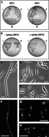
Inactivation of pyruvate kinase suppresses the ts1 mutant. (A) A transposon insertion in pyk restores viability to ts1 cells at 48°C. Strains were grown on tryptose blood agar plates at 30°C (permissive) or 48°C for 16 h. (i) Wild-type strain, SU110. (ii) ts1 (strain SU111). (iii) ts1 cells containing a suppressive transposon insertion in pyk. (B) Complementation of pyk restores the temperature-sensitive phenotype of ts1. Cells were grown in the presence or absence of 1% xylose for 16 h at 48°C. (i) Strain SU702 harboring the ts1 mutation and a deletion of the pyk gene. (ii) Strain SU610, in which pyk is expressed under xylose-inducible control at the ectopic amyE locus. (C to E) Deletion of pyk rescues ts1 cell division. Strains were grown in L broth for 1 h at 48°C and visualized by phase-contrast microscopy. (C) SU111 (ts1). (D) SU702 (ts1 Δpyk). The arrow shows an example of a gap separating divided cells. (E) SU110 (wild type). (F to H) pyk deletion rescues Z-ring formation in ts1. FtsZ localization was visualized by immunofluorescence microscopy after growth for 1 h at 48°C. (F) SU111 (ts1), showing no Z rings. (G) SU702 (ts1 Δpyk). Bright transverse bands represent normal-looking Z rings. (H) SU110 (wild type), showing normal Z rings at midcell. Scale bars, 5 µm.
FIG 2
FIG 2
Deletion of pyk affects Z-ring formation in B. subtilis. SU492 (amyE::Pxyl-ftsZ-yfp) and SU679 (Δpyk amyE::Pxyl-ftsZ-yfp) were grown to mid-exponential phase in the presence of 0.01% xylose at 37°C and visualized by phase-contrast and fluorescence microscopy. (A) SU492. (B) SU679. For both strains: i, phase-contrast image; ii, FtsZ-YFP localization, false colored in yellow; iii, DAPI staining of DNA, false colored in red; iv, overlay of panels i, ii, and iii. Arrows in panel Bi point to minicells. In panel Bii, filled arrows show examples of polar Z rings and the open arrow shows a cell with more than one ring. The arrow in Biv indicates a cell containing a polar Z ring and a single nucleoid with no visible evidence of segregation. The same cell is magnified in the inset. Scale bar, 5 µm.
FIG 3
FIG 3
Deletion of pyk renders B. subtilis hypersensitive to FtsZ overproduction. Panels A to E show wild-type and pyk mutant cells expressing various levels of FtsZ-YFP in addition to the native FtsZ protein. Strains were grown to mid-exponential phase at 37°C, and FtsZ was detected by fluorescence visualization of FtsZ-YFP (A to C) or by immunofluorescence microscopy (D to E). (A) SU679 (Δpyk amyE::Pxyl-ftsZ-yfp) expressing FtsZ-YFP in the presence of 0.2% xylose. Arrows indicate aberrant, irregular FtsZ structures. (B) SU679 cells in 0.01% xylose, showing a milder phenotype. (C) Control strain SU492 (amyE::Pxyl-ftsZ-yfp) showing normal midcell Z rings at the highest xylose concentration tested (0.2%). (D) Strain SU664 (Δpyk), lacking FtsZ-YFP altogether. Arrows point to polar Z rings. (E) Isogenic wild-type strain SU5, showing normal midcell Z rings. Scale bar, 5 µm. (F) Overproduction of untagged FtsZ is lethal in the pyk mutant background. Cells were grown in the presence or absence of 0.1 mM IPTG for 16 h at 37°C. (i) Wild-type strain SU5; (ii) SU558 (amyE::Pspachy-ftsZ); (iii) SU696 (Δpyk amyE::Pspachy-ftsZ).
FIG 4
FIG 4
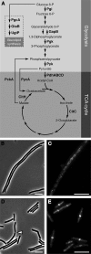
Effect of metabolic mutations on ts1 strain thermosensitivity. (A) Schematic representation of glycolysis, the TCA cycle, and the glucolipid biosynthesis pathway, highlighting 12 enzymes (bold) for which mutants were tested in this study. Pgi, phosphoglucose isomerase; GapB, glyceraldehyde-3-phosphate dehydrogenase; Pgk, phosphoglycerate kinase; Pyk, pyruvate kinase; PdhABCD, pyruvate dehydrogenase complex; CitC, isocitrate dehydrogenase; CitH, malate dehydrogenase; PckA, phosphoenolpyruvate carboxykinase; PycA, pyruvate carboxylase; PgcA, phosphoglucomutase; GtaB, uridine-diphosphoglucose pyrophosphorylase; UgtP, uridine-diphosphate glucosyltransferase. To assess the effect of metabolic mutations on the temperature-sensitive phenotype of ts1, each mutation was introduced into the ts1 genetic background, and the resulting strains (see Table S1 in the supplemental material) were grown for 1 h at 48°C. Cellular morphology was examined by phase-contrast microscopy, and FtsZ localization was visualized by immunofluorescence. (B) Phase-contrast image of strain SU703 (ΔcitC), shown as a representative of 11 mutants that did not suppress ts1. SU703 formed long, septumless filaments identical to those of normal ts1 cells (see Fig. 1C). (C) FtsZ localization in SU703 (ΔcitC), showing no Z rings. (D) Phase-contrast image of strain SU705, containing a suppressive mutation in pgk. SU705 cells were much shorter than those of the ts1 strain (7.5 ± 0.2 µm versus 33 ± 2 µm) and were often separated by clear gaps indicative of recently formed septa (one of these is highlighted by an arrow). (E) FtsZ localization in SU705, showing frequent Z rings. Scale bars, 5 µm.
FIG 5
FIG 5
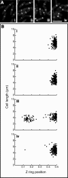
Addition of exogenous pyruvate rescues the Z-ring assembly defect of pyk mutant cells. SU492 (amyE::Pxyl-ftsZ-yfp) and SU679 (Δpyk amyE::Pxyl-ftsZ-yfp) were grown to mid-exponential phase at 37°C in the presence of 0.01% xylose and the presence or absence of 1% sodium pyruvate. FtsZ localization was visualized by fluorescence microscopy, and Z-ring positioning was measured to illustrate the rescue of Z rings back to the cell center in the pyk mutant. Z-ring position was defined as the distance from the ring to the nearest pole divided by the cell length, such that a value of 0.5 corresponds to midcell and a value of 0 to the cell pole. (A) Fluorescence micrographs of FtsZ localization. (B) Scatter plots of Z-ring positioning. For both panels A and B: i, SU492 without added pyruvate; ii, SU492 in 1% pyruvate; iii, SU679 without pyruvate; iv, SU679 in 1% pyruvate. Scale bar, 5 µm.
FIG 6
FIG 6
Localization of pyruvate dehydrogenase E1α in B. subtilis. Strains SU739 (amyE::Pxyl-pdhA-yfp) and SU742 (Δpyk amyE::Pxyl-pdhA-yfp) were grown to mid-exponential phase at 37°C in the presence of 0.1% xylose and the presence or absence of 1% sodium pyruvate. PDH E1α-YFP (encoded by the xylose-inducible pdhA-yfp fusion gene) was visualized in live cells using fluorescence microscopy. (A) SU739 in L broth without added pyruvate. (B) SU742 in L broth without pyruvate. (C) SU742 in L broth with 1% pyruvate added. (D) SU739 in Spizizen minimal medium (SMM); no added pyruvate. For panels A to D: i, phase-contrast image; ii, PDH E1α-YFP localization; iii, DAPI staining of DNA. Arrows provide reference points for comparing PDH E1α and nucleoid localization patterns within the same cell. Scale bar, 1 µm.
FIG 7
FIG 7
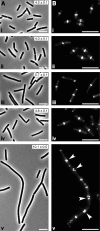
Depletion of PDH E1α affects Z-ring formation and cell division in a B. subtilis ezrA mutant. Cells were cultured in L broth to mid-exponential phase for 3 h at 37°C. Strains containing pdhA under xylose-inducible control were grown in the presence of 0.5% xylose to maintain pdhA expression or 1% glucose for maximal repression of the Pxyl promoter (83). It is important to note that 1% glucose or 0.5% xylose alone did not significantly affect division in wild-type cells (data not shown). Cellular morphology was examined by phase-contrast microscopy (A), and FtsZ localization was visualized by immunofluorescence (B). For both panels A and B: i, wild-type strain SU5; ii, strain SU791 (Pxyl-pdhA) supplemented with 1% glucose to deplete PDH E1α; iii, SU561 (ΔezrA); iv, SU792 (ΔezrA Pxyl-pdhA) in 0.5% xylose to maintain pdhA expression; v, SU792 (ΔezrA Pxyl-pdhA) with PDH E1α depleted. Arrows indicate helix-like localizations of FtsZ. Numbers represent average cell lengths ± standard errors of the means. Scale bar, 5 µm.
FIG 8
FIG 8
A nutrient-dependent role for PDH E1α in the control of Z-ring formation. Our results are consistent with a model in which PDH E1α (blue circles) acts as a positive regulator of FtsZ assembly and localizes over the nucleoid (gray) in a nutrient-dependent manner linked to pyruvate synthesis. PDH E1α exhibits only a weak association with the nucleoid under minimal-nutrient conditions (left), while in nutrient-rich media (center), it localizes much more strongly over the chromosome. Importantly, the negative regulatory systems Min and nucleoid occlusion (NO) are known to inhibit FtsZ polymerization at locations other than the cell center, and this could help to restrict the influence of PDH E1α on Z-ring formation to midcell. During the late stages of chromosome segregation (all cells are depicted at this point in the cell cycle), the proteins that mediate nucleoid occlusion are absent from the central region of the cell due to a lack of Noc/SlmA binding sites around the chromosome terminus. The amount of PDH E1α located within this central region increases with rising nutrient levels, and this could provide a positive signal for Z-ring formation at midcell that becomes stronger under nutrient-rich conditions (in which cells grow faster and must therefore divide more frequently). When pyruvate synthesis is artificially blocked in a pyk mutant, PDH E1α fails to colocalize with the nucleoid at all (right). Accumulation of PDH E1α at the nucleoid-free cell poles under these conditions could trigger polar Z-ring formation by overcoming the inhibitory effects of the Min system to generate a net positive signal for FtsZ assembly.

Similar articles

Cited by

References

    1. Harry E, Monahan L, Thompson L. 2006. Bacterial cell division: the mechanism and its precision, p 27–94 In Jeon KW, International review of cytology: a survey of cell biology, vol. 253 Academic Press, San Diego, CA. - PubMed
    1. Adams DW, Errington J. 2009. Bacterial cell division: assembly, maintenance and disassembly of the Z ring. Nat. Rev. Microbiol. 7:642–653. 10.1038/nrmicro2198 - DOI - PubMed
    1. de Boer PA. 2010. Advances in understanding E. coli cell fission. Curr. Opin. Microbiol. 13:730–737. 10.1016/j.mib.2010.09.015 - DOI - PMC - PubMed
    1. Erickson HP, Anderson DE, Osawa M. 2010. FtsZ in bacterial cytokinesis: cytoskeleton and force generator all in one. Microbiol. Mol. Biol. Rev. 74:504–528. 10.1128/MMBR.00021-10 - DOI - PMC - PubMed
    1. Osawa M, Anderson DE, Erickson HP. 2008. Reconstitution of contractile FtsZ rings in liposomes. Science 320:792–794. 10.1126/science.1154520 - DOI - PMC - PubMed

Publication types